Section: Abdominal wall and retroperitoneum Curriculum: Curriculum, page 3
Embryology
- Abdominal wall forms 4/40 causing infolding in the craniocaudal and mediolateral directions
- Lateral abdominal folds of embryo meet in anterior midline and surround the yolk sac
- 6/40 - rapid growth of intestines - herniation of midgut into umbilical cord.
- Cranial limb - lengthens ++
- Caudal limb lengthens ++
- 6-10/40 - elongation and rotation of midgut
- During herniation, midgut rotates 90 degrees counter clockwise
- 10/40 - return of gut into abdomen - duodenum, ascending colon and descending colon fixed retroperitoneally
- As the gut returns to the abdomen, it rotates another 180 degrees counterclockwise - thus the ascending colon and caecum is to the right of the small bowel, the descending colon is to the left, and the DJ flexure to the left of the SMA.
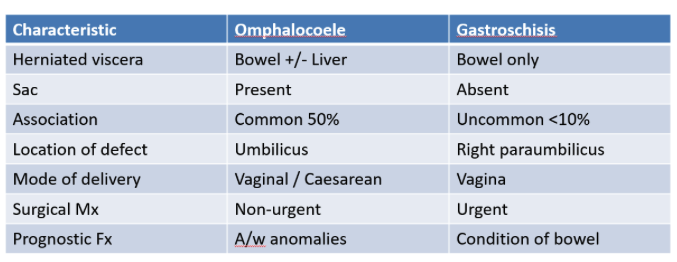
Gastroschisis
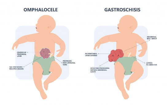
- Definition: Full thickness paraumbilical abdominal wall defect associated with evisceration of bowel defect associated with evisceration of bowel.
- Usually right paraumbilical area.
- Usually < 4cm.
- Abdominal wall defect to the right of the cord insertion
Aetiology
- Not clear - below are some possible explanations
- Failure of mesoderm to form in anterior abdominal wall.
- Failure of lateral folds to fuse in midline - leaving defect at right of umbilicus.
- Thrombosis of right umbilical vein causing local necrosis of abdominal wall
- Is associated with intestinal atresia
- Increased risk with maternal use of vasoconstrictive drugs/smoking.
Omphalocoele
- Displacement of abdominal contents into the umbilical cord
- Unlike umbilical hernia - omphalocoele only covered with peritoneum.
- Cause: improper retraction of the physiologic umbilical hernia during foetal development - therefor no proper muscle formation could take place in this region.
- Other intra-abdominal viscera (liver, bladder, stomach, gonads) can also be found within the sac.
Surgical management
- Goal: Return of viscera into abdominal cavity while minimising risk of damage OR abdominal compartment syndrome.
- At time of delivery - tertiary care center, covering the defect with gauze dressings soaked in thermally neutral sterile, covering the dressing with clear plastic wrap, inserting an orogastric tube to decompress the stomach, stabilizing the airway to ensure adequate ventilation
- Omphalocoele
- Worse outcomes overall but this is due to association with other congenital defects
- Small defect can be closed in first 24-72 hours
- If not possible then staged closure
- Gastroschisis
- Managed operatively and closed within a few hours of birth, if possible.
- Should there not be enough domain, a silo can be placed to stop the abdominal wall contracting and staged closure be performed.