Section: Abdominal wall and retroperitoneum Sub-section: Hernias Curriculum: Curriculum, page 2
Definition
- Protrusion of abdominal contents through a defect in the musculoaponeurotic layers of a surgical (abdominal) scar
Incidence
Rates after laparotomy: 10-20%
Clinical
- Can develop as early as 1-month post-op
- Usually occur within the first year after surgery, but may occur later than this
- C.f. Dehiscence:
- Develops at 5-10 days
- +/- preceded by serosanguinous discharge from the wound
Aetiology
- Patient factors
- Obesity
- Age
- Nutrition
- Immunosuppression: DM , steroids/cytotoxicity
- Wound infection Post-op
- Post-op pulmonary complications / vomiting
- Technical factors
- Poor surgical technique
- Ideal = Sutures 1cm apart, total length > 4x the length of the wound
- Placement of drains & stomas in the primary operative wound
- Failure to close the fascia of a laparoscopic trocar > 10mm in size
- ? fewer incisional hernias after transverse vs midline incision
Management
- Repair small defects
- Large defects can be left if asymptomatic
- Unlikely to incarcerate
- Pre-op optimisation
- Weight loss, stop smoking etc
- Technique
- Primary repair – not recommended, recurrence 50-60%
- Open mesh – recurrence rates 30%
- Lap mesh repair – gold standard for large ventral hernias
- Component separation if large defect
Open Mesh Repair
- 3cm overlap recommended (as the mesh shrinks)
- Types of mesh repair:
- Inlay
- Sublay / underlay / preperitoneal / “retro-rectus” placement
- Mesh between peritoneum & fascia (or peritoneum & muscle)
- Lowest recurrence rate
- ? fewer complications
- Onlay with primary closure
- Mesh superficial to rectus, after closing rectus
- European Hernia society guidelines
- Recommend retromuscular (retrorectus or pre-peritoneal)
- Onlay vs retrorectus
- Onlay - increased risk of recurrence, seroma and haematoma
- Low quality evidence
- Onlay - increased risk of recurrence, seroma and haematoma
- Onlay vs retrorectus
- Recommend against IPOM if possible to limit contact with viscera
- Recommend retromuscular (retrorectus or pre-peritoneal)
Laparoscopic Ventral Hernia Repair
IPOM
- Supine, arms out, iodine drape
- Ports
- 3 x 5mm ports as lateral as possible on one side, after safe pneumoperitoneum
- Left subcostal if no previous surgery in this area
- 3 x 5mm ports as lateral as possible on one side, after safe pneumoperitoneum
- Dissection
- Sharp/blunt dissection of adhesions
- Inspect bowl immediately if close to r/o injury
- Adhesions to the sac – can divide sac and leave it on bowel, apply external pressure to the sac
- Post-adhesiolysis
- Use spinal needle from outside to delineate edge of hernia
- Mark on the outside
- Decompress pneumoperitoneum and mark 3cm out from edge of hernia
- Take mesh (covered mesh) and cut to marked size on abdominal wall
- Mark mesh for orientation
- Mesh placement
- 2x further 5mm ports on opposite side
- Place 4 x sutures (0 Prolene) to corners and then place into abdomen with smooth side down
- Bring pre-placed sutures out at pre marked area (scalpel incision at this spot)
- Tie knots down on to fascia, use local here as can be sore
- Reduce insufflation pressure to 10mmHg
- Absorbitac around the edge of the mesh + sutures (with suture passing device) every few cm
- Close 5mm ports
Pre-peritoneal
- Similar to above however raise a flap of peritoneum
- Dissection around the sac reducing it down
- Place mesh
- Close peritoneum over the top
Prognosis/Natural Hx
- 50% are apparent at 1yr post-op
- Natural Hx:
- Incarceration in 15%; strangulation in 2%
- Recurrence rates after repair
- After primary re-suture: ≈ 25-46%
- After open mesh repair: 10-20%
- 19% after onlay mesh & 7.3% after sublay mesh
- After lap mesh repair ≤ 10% (1-16%)
- Low recurrence rates ?due to
- ↓ infection (due to no subcut dissection)
- No drains
- ↑ pickup of other hernial defects
- Recurrence after repair of a recurrent incisional hernia:
-
50%
- 60-90% of recurrences occur within 2yrs
-
Loss of Domain
- International Consensus Definition 2020:
- “A ventral hernia large enough such that simple reduction in its contents and primary fascial closure either cannot be achieved without additional reconstructive techniques OR cannot be achieved without significant risk of complications due to the raised intra-abdominal pressure”
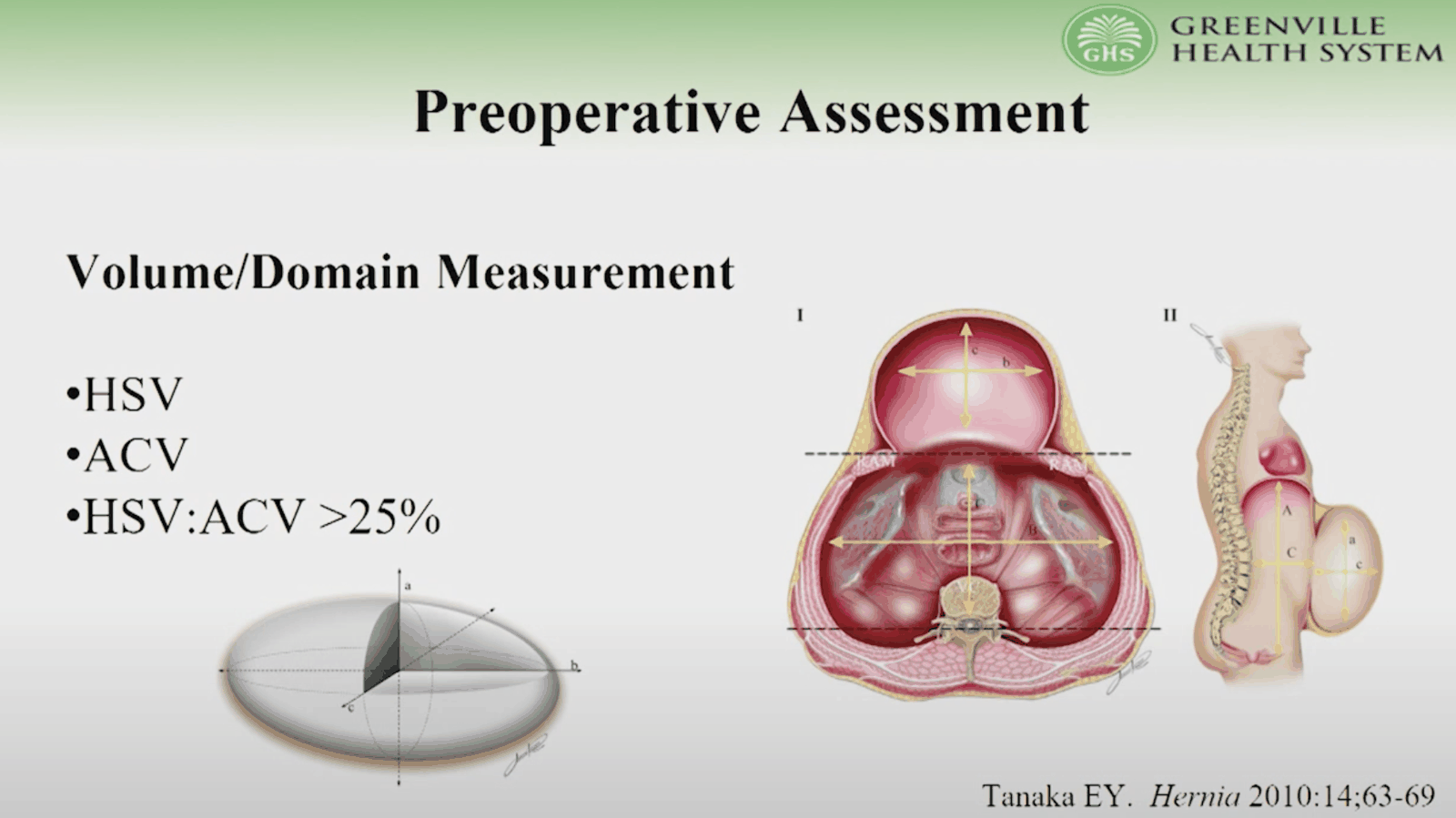 If ratio >25% then LOD and need for techniques such as botox or progressive pneumoperitoneum
Component seperation
- Options
- Non-surgical
- Using pre-operative Botox
- Surgical
- Posterior rectus release (Rives Stoppa)
- External oblique release
- Transverse Abdominis release
- Non-surgical
Posterior rectus release (Rives Stoppa)
- First step in the bottom two
- Create a retro rectus space
- If there is enough movement then can close posterior sheath, place mesh and close anterior sheath
External oblique release
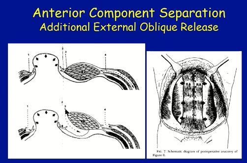
- Rectrorectus mobilisation and then anterior release of external oblique
Transverse Abdominis release
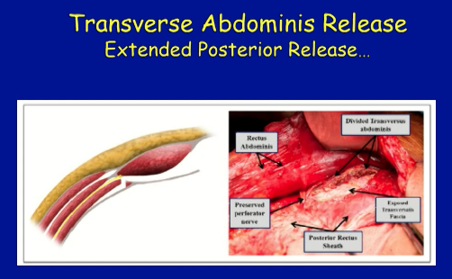
- Continue from rectorectus plane and divide transverse abdominus muscle
- To create a larger plane for mesh placement and also more medialiastion of sheath