Peritoneal Compartments
- Peritoneal cavity divided into compartments
- Supracolic
- Infracolic
- Pelvic
- Dividing line between Supracolic and Infracolic compartments
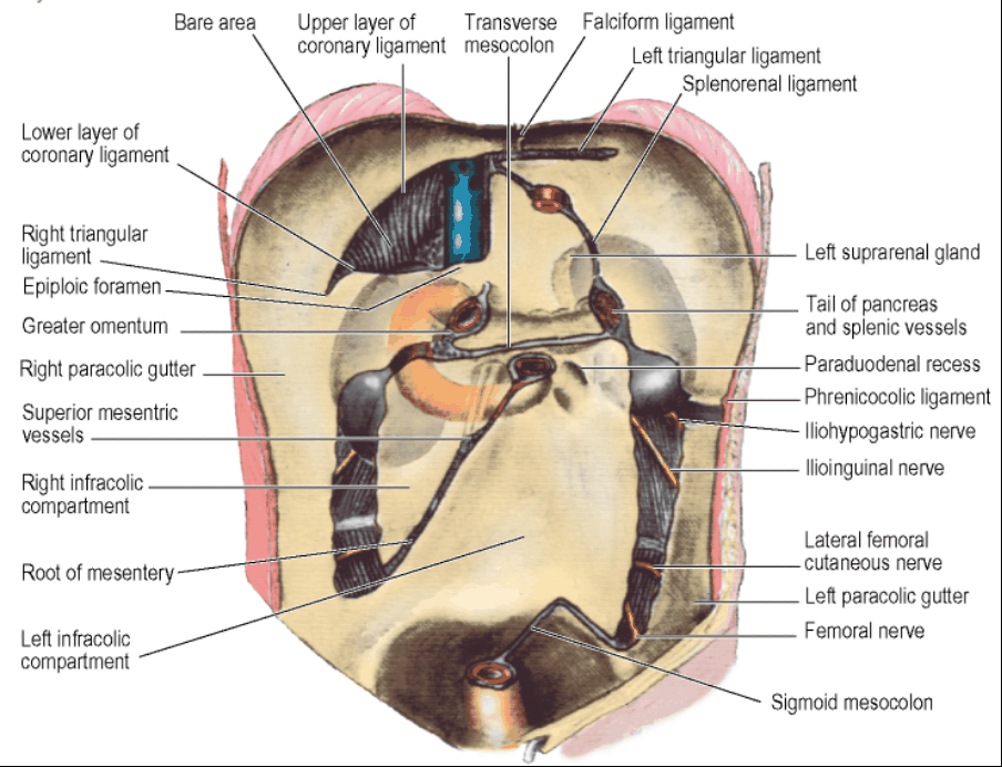
- Attachment of the Transverse Mesocolon to the posterior abdominal wall
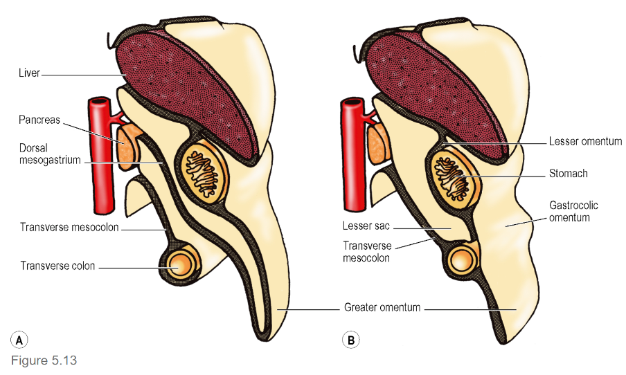
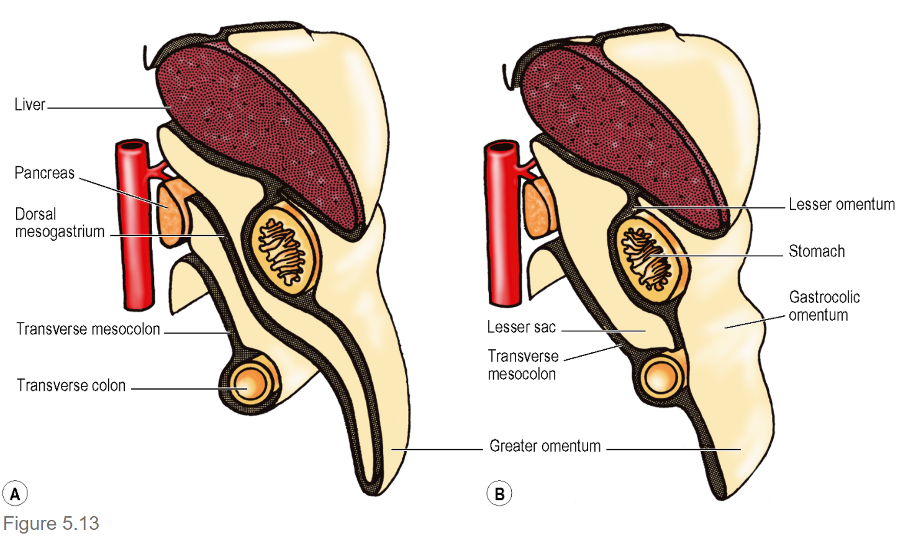
Lesser Sac
- A.k.a. Omental Bursa
- Opens into the Greater Sac through the Epiploic Foramen
Relationships
- Anterior Wall:
- Posterior layer of Lesser Omentum
- Peritoneum over the posterior aspect of the Stomach
- Posterior of the two anterior layers of the Greater Omentum
- Posterior Wall:
- Anterior of the two posterior layers of the Greater Omentum
- Adheres to but is surgically separable from the anterior surface of the Transverse Colon and Transverse Mesocolon
- Peritoneum that covers:
- Front of the neck and body of Pancreas
- Upper part of Left Kidney
- Left Suprarenal Gland
- Commencement of Abdominal Aorta
- Coeliac Artery (Plexus and Nodes)
- Part of the Diaphragm
- NB: Common Hepatic Artery also in the posterior wall
- Anterior of the two posterior layers of the Greater Omentum
- Upper border
- Narrow and located at the right side of the abdominal Oesophagus
- Where the peritoneum of the posterior wall is reflected anteriorly on the inferior aspect of the diaphragm to form the posterior layer of the Lesser Omentum
- Lower border
- Theoretically can extend down between the anterior two layers and the posterior two layers of the Greater Omentum
- However, fusion of the two layers of the Greater Omentum means the lesser sac does not extend much below the Transverse Colon
- Lesser sac extends behind the first 2.5cm of Duodenum
- Theoretically can extend down between the anterior two layers and the posterior two layers of the Greater Omentum
- Left border
- Formed by the Splenorenal and Gastrosplenic ligaments at the tail end of the Pancreas
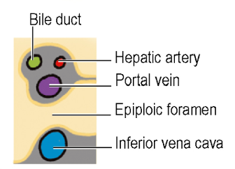
Epiploic Foramen (of Winslow)
- A vertical slit about 2.5cm at the right border of the Lesser Sac
- Boundaries:
- Upper:
- Caudate Process of the Liver
- Lower:
- 1st part of the Duodenum
- Posterior:
- IVC covered by parietal peritoneum of the posterior abdominal wall which, continuing to the left through the foramen, becomes the peritoneum of the posterior wall of the lesser sac
- Anterior:
- Bounded by the right free margin of the Lesser Omentum
- Containing between its two layers the Portal Vein and (anterior to it) the Hepatic Artery and Bile Duct (Duct to the right of the Artery)
- as well as autonomic nerves, lymphatics and nodes
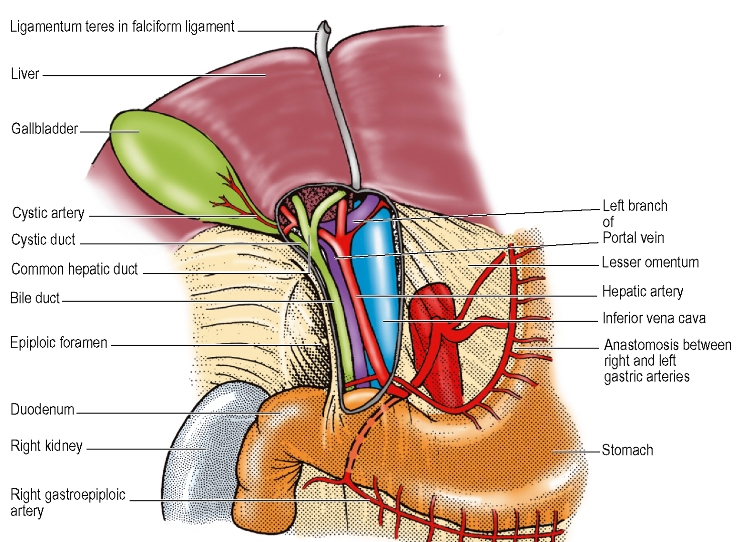
- as well as autonomic nerves, lymphatics and nodes
- Containing between its two layers the Portal Vein and (anterior to it) the Hepatic Artery and Bile Duct (Duct to the right of the Artery)
- Bounded by the right free margin of the Lesser Omentum
- Upper:
Lesser Omentum
-
Definition
- Two layers of peritoneum extending between the Liver and the Lesser Curvature of the Stomach
- A.k.a. Gastrohepatic Omentum
- Two layers of peritoneum extending between the Liver and the Lesser Curvature of the Stomach
-
Attachments
- Extends from right side of the abdominal Oesophagus and along the Lesser Curvature to the first 2cm of the Duodenum
- Liver attachment is ‘L’ Shaped to the Fissure for the Ligamentum Venosum and the Porta Hepatis.
- NB: There is NO attachment to the fissure for the Ligamentum Teres
-
Superior extension
- Two layers enclose the Liver and then spread onto the diaphragm and anterior abdominal wall as the Coronary, Triangular and Falciform Ligaments
- Anterior layer continuous with the posterior layer of the Left Triangular Ligament
- Two layers enclose the Liver and then spread onto the diaphragm and anterior abdominal wall as the Coronary, Triangular and Falciform Ligaments
-
Inferior extension – Two layers become the Greater Omentum
-
NB: Derived from the Ventral Mesogastrium
Transverse Mesocolon
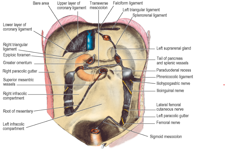
- Double fold of peritoneum
- Passes from Transverse Colon to the front of the 2nd part of the Duodenum and to front of Head and Body of the Pancreas
-
Adherent to the posterior surface of the Greater Omentum
-
Supracolic Compartment
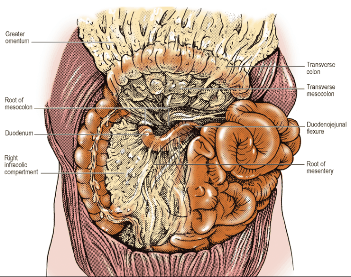
- Attachments of Liver to the Diaphragm and abdominal wall define the subdivisions of the Supracolic compartment
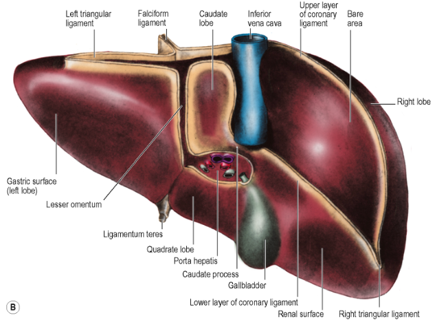
- Falciform Ligament – divides the Left and Right Subphrenic (Subdiaphragmatic) spaces
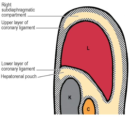
- Two spaces closed above by the superior layer of the Coronary Ligament and the anterior layer of the Left Triangular Ligament
- Right Subhepatic Space (Hepatorenal Pouch of Morrison)
- Behind the right lobe of the Liver and in front of the Right Kidney
- Superiorly, space closed above by the inferior layer of the Coronary Ligament and the small right Triangular Ligament
- To the right, bounded by the abdominal surface of the Diaphragm
- To the left, the space communicates through the Epiploic Foramen with the Lesser Sac or Left Subhepatic Space
- Inferiorly, it is continuous with the Right Paracolic Gutter
- When supine, Hepatorenal pouch is the lowest part of the peritoneal cavity (with the sole exception of the pelvis)
- Thus, area of intraperitoneal fluid accumulation
Infracolic Compartment
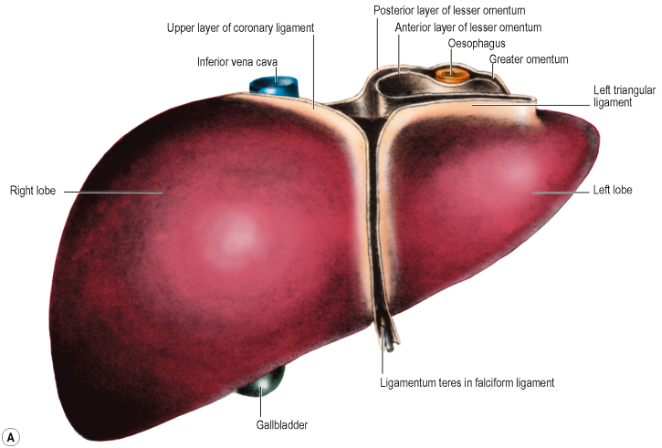
- Divided into two by the attachment of the Root of the Mesentery of the Small Bowel
- To the right of the Mesentery: Right Infracolic space
- Triangular
- Apex at the Ileocaecal Junction
- Right side: Ascending Colon
- Upper border: Attachment of the Transverse Mesocolon
- To the left of the Mesentery: Left Infracolic space
- Quadrilateral
- Widens below where it is continuous across the Pelvic Brim with the pelvic cavity
- Left side: Descending Colon
- Upper border: Attachment of the Transverse Mesocolon
Small Bowel Mesentery
- Course: Passes down from left to right at an angle of about 45°, begins at the Duodenojejunal Junction ⇒ Crosses 3rd part of the Duodenum where the Superior Mesenteric Vessels enter between its two layers ⇒ Continues downwards across the Aorta, Inferior Vena Cava, Right Psoas Muscle, Right Ureter ⇒ Reaches Right Iliac Fossa
- NB: Root of the Mesentery also related posteriorly to the Right Gonadal Vessels
- Length of attachment: 15cm
- Depth of mesentery (from root to gut): 20cm at its deepest (i.e. in the central part)
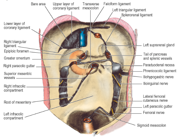
- Numerous Pacinian Corpuscles are located in the retroperitoneal tissue in the region of the Mesentery Root – tension and traction on peritoneal folds in the upper abdo cause a drop in blood pressure by undue stimulation of these encapsulated mechanoreceptors
Right Paracolic Gutter
- Lateral to the Ascending Colon
- Traced upwards into the Hepatorenal Pouch
- Traced downwards into the pelvis
Left Paracolic Gutter
- Lateral to the Descending Colon
- Limited above by the Phrenicocolic Ligament (small transverse fold of peritoneum between the Splenic Flexure and Diaphragm
- Traced downwards – left attachment of the lateral limb of the Sigmoid Mesocolon at the Pelvic Brim
Sigmoid Mesocolon
- Upside-down V-Shape
- Two limbs diverge from each other at the bifurcation of the Common Iliac Vessels on the Pelvic Brim over the Left Sacroiliac Joint
- Left Ureter lies over the bifurcation of the Common Iliac Artery
- Inferior Mesenteric Vessels lie medial to the Left Ureter
- Inferior Mesenteric Vein lies between the Inferior Mesenteric Artery and the Left Ureter
- Lateral limb
- Passes forwards along the Pelvic Brim over the External Iliac vessels halfway to the Inguinal Ligament
- Medial limb
- Slopes down into the hollow of the Sacrum
- Reaches the midline in front of S3 at the commencement of the Rectum
Nerve Supply of Peritoneum
- Partial Peritoneum supplied segmentally by spinal nerves innervating adjacent muscles
- Diaphragmatic Peritoneum supplied centrally by the Phrenic Nerve (C4)
- Remainder of Parietal Peritoneum supplied segmentally by Intercostal and Lumbar Nerves
- In pelvis, Obturator nerve is the chief source of supply
- Visceral Peritoneum – innervated by afferent nerves which travel with the autonomic supply to the viscera
- Pain from diseased viscera due to ischaemic, muscle spasm and stretching of the visceral peritoneum (including mesenteric folds) or involvement of the parietal peritoneum
Retroperitoneal Space
- Structures on posterior abdominal wall behind peritoneum:
- Aorta
- Inferior Vena Cava
- Cisterna Chyli
- Lymph nodes and lymphatics
- Nerves – mostly branches of the Lumbar Plexus, including Sympathetic Trunks
- Kidneys
- Ureters
- Pancreas
- Ascending and Descending Colon
- Duodenum (most)
- Suprarenal Glands