Overview
- Lie anterosuperior to the upper part of each kidney
- Lie within their own compartment of the Renal Fascia
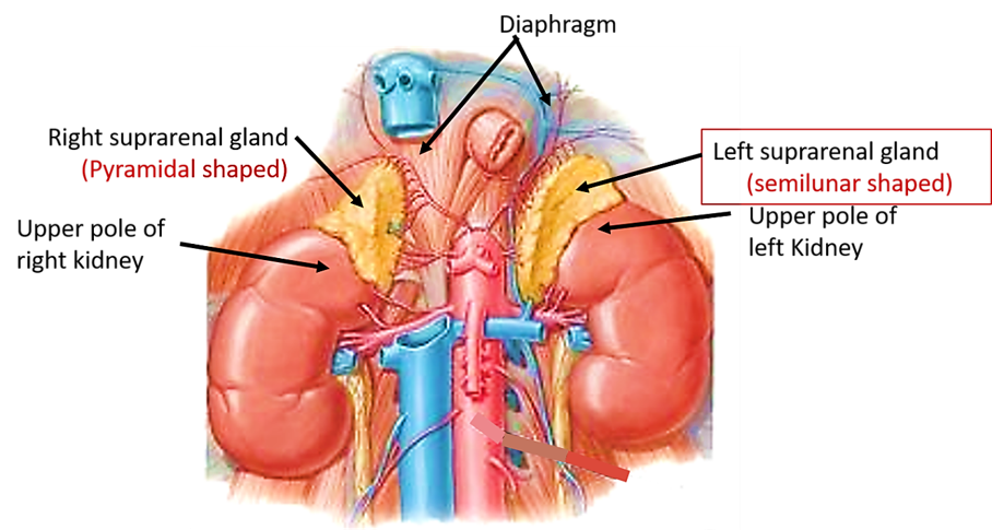
Right Suprarenal Gland
- Shape: Pyramidal
- Lies on the Diaphragm (NB: Right Crus)
- Encroaches onto the front of the Right Kidney
- Anterior surface
- Overlapped medially by the Inferior Vena Cava
- Rest of anterior surface in contact above with the Bare Area of the Liver
- Covered below by Peritoneum of the posterior wall of the Hepatorenal Pouch
Left Suprarenal Gland
- Shape: Crescentic
- Drapes over the medial border of the Left Kidney above the Hilum
- Lies on the Left Crus of the Diaphragm
- Overlaps the front of the Left Kidney
- Anterior surface:
- Upper part covered by Peritoneum of the posterior wall of the Lesser Sac forming part of the Stomach Bed
- Lower part – in contact with the body of the Pancreas and the Splenic Vessels
Blood Supply
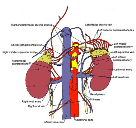
- 3 Sources
- Directly from the Aorta
- Renal Artery
- Inferior Phrenic Artery
- Venous Drainage
- Left - Adrenal Vein to Left Renal Vein
- Right – Adrenal Vein to IVC (short)
- Veins drain Adrenal Medulla
- Cortical drainage through medulla
Lymphatic Drainage
- To Para-aortic Lymph Nodes
Nerve Supply
- Medulla
- Main supply – myelinated pre-ganglionic sympathetic fibres from the Splanchnic nerves via the Coeliac Plexus
- Synapse directly with Medullary Cells
- Blood vessels receive usual Post-Ganglionic Sympathetic fibres
- Richly supplied by Splanchnic Fibres (T5-9)
- Can be regarded as modified sympathetic ganglion
- Axon replaced by secretory cells
- Chromaffin cells/phaeochromcytes
- Filled with granules containing catecholamines
- Axon replaced by secretory cells
- Can be regarded as modified sympathetic ganglion
- Main supply – myelinated pre-ganglionic sympathetic fibres from the Splanchnic nerves via the Coeliac Plexus
- Cortical
- Hormone driven e.g. ACTH from the Anterior Pituitary Gland causes release of cortisol
Structure
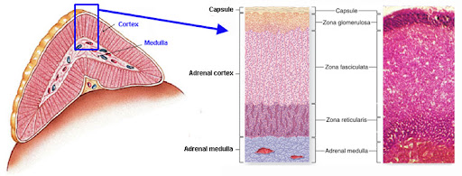
- Outer yellow cortex enclosing thinner grey medulla
- Cortex
- Produces cortisol, aldosterone, androgens and related hormones
- 3 layers/zones
- Zona Glomerulosa – small, rounded cells
- Zona Fasciculata – parallel rows of pale-staining vacuolated cells
- Zona Reticularis – smaller and darker-staining cells
- Medulla
- Larger cells secreting catecholamines adrenaline (epinephrine) (80%) and noradrenaline (norepinephrine) (20%) and some dopamine
- Many medullary cells exhibit chromaffin reaction – contain fine cytoplasmic granules (catecholamine precursors) which are coloured brown by chromium salts
- Dilated capillaries prominent (unlike the cortex)
- NB: Contains cells equivalent to post-ganglionic neurones
Microscopic Anatomy
Zona Glomerulosa
- Columnar cells, relatively little cytoplasm compared to nuclei, organised in clusters
- Produces mineralocorticoids
Zona Fasciculata – (75% of Cortex)
- Poorly staining polyhedral cells, organised in radial columns
- Primarily produces glucocorticoids
Zone Reticularis
- Rounded branching cords of cells
- Produce sex hormones, particularly DHEAS
Medulla – 15% volume
- Vascular spaces
- Eosinophilic phaeochromocytes with polymorphic nuclei
- Uptake dichromate – “Chromaffin cells”
Surgical Approach
- Bilateral adrenalectomy
- Glands approached from the front
- Anterior transperitoneal approach also used for laparoscopic bilateral adrenalectomy
- Bilateral subcostal rooftop incision
- Transperitoneal access
- Exposing Right Suprarenal Gland:
- Reaction of Right Lobe of Liver
- Kocher Manoeuvre to mobilize 2nd part of Duodenum and Head of Pancreas from the upper pole of the Kidney and Inferior Vena Cava
- Exposing the Left Suprarenal Gland:
- Divide Phrenicocolic Ligament to mobilize Splenic Flexure of the Colon
- Posterior layer of the Splenorenal Ligament incised
- Spleen turned medially with the Tail of the Pancreas
- On each side – Adrenal Vein ligated before the numerous small arteries
- Right vein particularly short and Inferior Vena Cava easily torn
- Glands must be handled as little as possible before venous ligation to prevent surges of hormone release
- Posterior extraperitoneal approach
- Through bed of 12th Rib
- Provides access for unilateral adrenalectomy
- Removal of a large adrenal tumour may require a higher approach – through the bed of the 11th or 10th Ribs and division of the Diaphragm