Overview
- C-shaped
- Length: 25cm
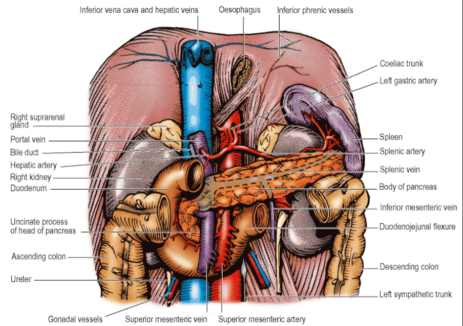
- Anterior and to the right of the Inferior Vena Cava and Aorta
- First 2.5cm are contained between the peritoneum of the Lesser and Greater Omenta
- Remainder is retroperitoneal
- Divided into 4 parts – superior, descending, horizontal and ascending or 1st, 2nd, 3rd and 4th
- Lengths: 2, 3, 4, and 1 inches (or 5, 7.5, 10 and 2.5cm)
- C-shaped loop around the Head of the Pancreas
- Opposite body of L2 vertebra
- 1st part – Lie at the level of L1
- 2nd part – Right side of L2
- 3rd part – Crosses in front of L3
- 4th part – Left side of L2
Summary
- First (Superior) Part:
- Extends from the pylorus of the stomach to the superior duodenal flexure.
- Lies at the level of L1.
- Second (Descending) Part:
- Extends from the superior duodenal flexure to the inferior duodenal flexure.
- Passes anterior to the hilum of the right kidney.
- Contains the openings of the common bile duct and pancreatic duct.
- Third (Horizontal) Part:
- Extends from the inferior duodenal flexure across the midline at L3.
- Passes posterior to the superior mesenteric vessels.
- Fourth (Ascending) Part:
-
Extends from L3 to the duodenojejunal flexure, supported by the ligament of Treitz.
-
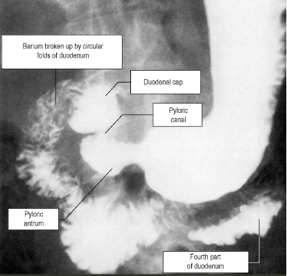
- Mucous membrane thrown into numerous circular folds (Plicae Circulares or Valvulae Conniventes)
- Walls of the first 2.5cm are smooth – hence smooth outline of the full shadow of barium in the Duodenal Cap on x-ray
First Part of the Duodenum
- Course: Runs to the right, upwards, and backwards from the Pylorus
- First 2.5cm
- The Duodenal Cap
- Lies between the peritoneal folds of the Greater and Lesser Omenta
- Forms the lowermost boundary of the opening into the Lesser Sac
- Relations of first 2.5cm (Duodenal Cap):
- Anterior to
- Gastroduodenal Artery
- Bile Duct
- Portal Vein
- Inferior Vena Cava
- NB: In part of its course, the 1st part of the Duodenum is in contact with the anterior surface of the Inferior Vena Cava (Part 1 Bank)
- Posterior to
- Gallbladder
- Anterior to
- The Duodenal Cap
- Relations of second 2.5cm
- Courses backwards and upwards on the upper part of the Head of the Pancreas to the medial border of the Right Kidney
- NB: The 1st part of the Duodenum is above the hilum of the Right Kidney
- Covered in front with peritoneum
- Right lobe of Liver lies over this peritoneum
- Posterior surface
- Bare of peritoneum
- Courses backwards and upwards on the upper part of the Head of the Pancreas to the medial border of the Right Kidney
Second Part of the Duodenum
- Course
- Curves downwards over the hilum of the Right Kidney
- Relation
- Anterior
- Covered in front with peritoneum
- Crossed by the attachment of the Transverse Mesocolon
- Anterior
- Upper half lies in the Supracolic compartment to the left of the Hepatorenal Pouch (in contact with the Liver)
- Lower half lies in the right Infracolic compartment medial to the inferior pole of the Right Kidney (in contact with coils of jejunum)
- Lies alongside the Head of the Pancreas
- Posteromedial wall receives the common opening of the Bile Duct and Main Pancreatic Duct at the Hepatopancreatic Ampulla (of Vater) – opens on the summit of the Major Duodenal Papilla
- Halfway along the second part, 10cm from the pylorus
- Overlapped by semilunar flap of mucous membrane
- Small opening of the Accessory Pancreatic Duct (on the Minor Duodenal Papilla) lies 2cm proximal to this opening
- NB: Junction of 2nd and 3rd part of Duodenum directly related to the Right/Ascending Colon
Third Part of the Duodenum
- Course: Curves forwards from the right Paravertebral Gutter over the slope of the Right Psoas muscle - Gonadal vessels and Ureter intervening - Passes over the forwardly projecting Inferior Vena Cava and Aorta to reach the Left Psoas muscle
- As inferior border crosses Aorta, it lies on the commencement of the Inferior Mesenteric Artery
- Upper border hugs the lower border of the Pancreas
- Covered by the peritoneum of the posterior abdominal wall just below the Transverse Mesocolon
- Crossed by the Superior Mesenteric Vessels and by the leaves of the commencement of the mesentery of the Small Intestine sloping down from the Duodenojejunal Flexure
- Thus, lies in both the right (mainly) and left Infracolic compartments
- Anterior surface in contact with coils of Jejunum
Fourth Part of the Duodenum
- Course:
- Ascends to the left of the Aorta
- Lies on the left Psoas muscle and left Lumbar Sympathetic Trunk
- Reaches the lower border of the Body of the Pancreas
- Covered in front by the peritoneal floor of the left Infracolic compartment by coils of jejunum
- Breaks free from the peritoneum that plastered it down to the posterior abdominal wall and curves forwards and to the right as the Duodenojejunal Flexure
- Pulls up a double sheet of peritoneum from the posterior abdominal wall, the mesentery of the small intestine, which slopes down to the right across the Third Part of the Duodenum and posterior abdominal wall
- NB: The 4th Part of the Duodenum does not have its own mesentery
- Pulls up a double sheet of peritoneum from the posterior abdominal wall, the mesentery of the small intestine, which slopes down to the right across the Third Part of the Duodenum and posterior abdominal wall
- Duodenojejunal flexure fixed to the Left Psoas Fascia by fibrous tissue
- Further supported by the Suspensory Muscle of the Duodenum (Muscle or Ligament of Trietz)
- Thin band of connective tissue which may contain muscle – skeletal musclen fibres from the Right Crus of the Diaphragm to connective tissue around the Coeliac Trunk and smooth muscle fibres that run from there, behind the Pancreas and in front of the Left Renal Vein, to the muscle coat of the flexure.
- Further supported by the Suspensory Muscle of the Duodenum (Muscle or Ligament of Trietz)
Paraduodenal Recesses
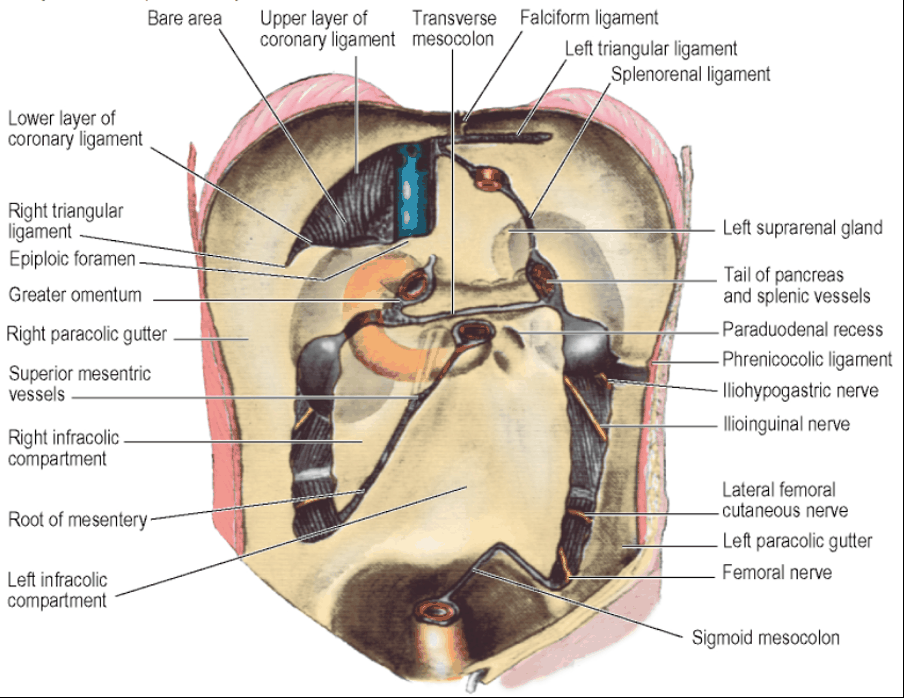
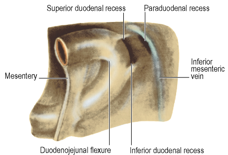
- To the left of the Duodenojejunal Flexure – certain peritoneal folds may cover recesses or fossae
- Paraduodenal Recess Proper – lies behind a fold raised by the upper end of the Inferior Mesenteric Vein (incarcerated internal hernia in this fossa may obstruct and thrombose the vein)
- Horizontal folds of peritoneum may cover Superior and Inferior Duodenal Recesses
- Retroduodenal recess may be found behind the flexure
Blood Supply
- Arterial Supply: Superior and Inferior Pancreaticoduodenal Arteries
- First 2cm – receives blood from the Hepatic, Gastroduodenal, Supraduodenal, Right Gastric and Right Gastroepiploic Arteries
- Venous drainage: Tributaries of the Superior Mesenteric and Portal Veins
Lymph Drainage
- Channels that accompany the Superior and Inferior Pancreaticoduodenal Vessels to the Coeliac and Superior Mesenteric Nodes
Duodenal Ulcers
- Bleeding Duodenal Ulcer commonly located in posterior aspect of the first 2cm of the Duodenum
- The Gastroduodenal Artery is the vessel it commonly erodes into