Biliary tract
Normal
- Right and left leave liver
- Becomes common hepatic duct
- At the point that the cystic duct join then becomes CBD
- CBD 3 parts
- Supra-duodenal - above the duo
- Retro-duodenal - behind
- Para-duodenal - groove at back of head of pancreas
60% have normal anatomy
Right
- 5, 6, 7 and 8 + 1
- Short extra-hepatic compared to left
- Right posterior sectoral duct (6,7)
Left
- 2,3,4 + 1
- Longer extra-hepatic
Couinaud (Blumgart) classification
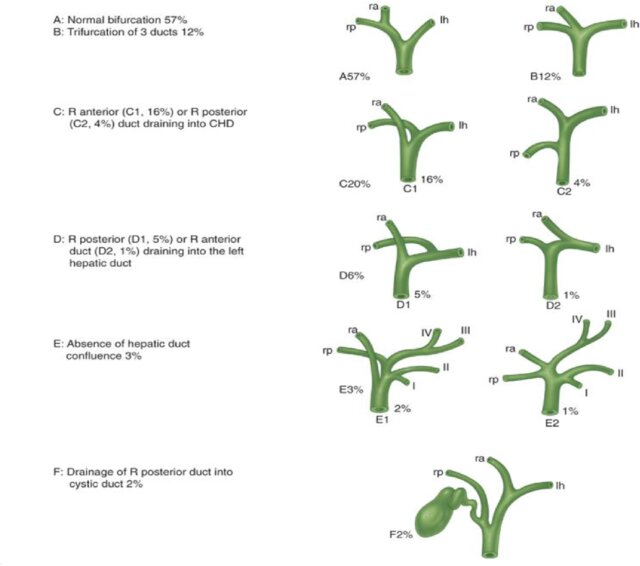 A- normal (60%)
B - trifurcation of 3 ducts
C - right sectoral into CHD
D - right sectoral into left hepatic
E - Abscence of hepatic duct
F - Right posterior sectoral duct into cystic duct
A- normal (60%)
B - trifurcation of 3 ducts
C - right sectoral into CHD
D - right sectoral into left hepatic
E - Abscence of hepatic duct
F - Right posterior sectoral duct into cystic duct
Arterial supply of CBD
- Right hepatic artery - hilar
- Gastroduodenal artery
- Pancreaticoduodenal arcades - distal
- Axial 3, 9 o’clock position
Duct of Luschka
- Subvesical duct which lies within the gallbladder bed of the liver close to the surface and can be damaged during removal of the posterior gallbladder wall.
Cystic artery
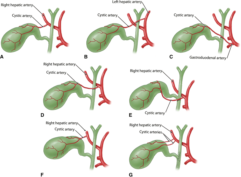 A - indicates the cystic artery originating from right hepatic artery or aberrant right hepatic artery.
B- shows the cystic artery originating from left hepatic artery
C - from the gastroduodenal artery.
D - traveling anterior to common hepatic duct
E - travels anterior to common bile duct and inferior to cystic duct.
F - short cystic artery is seen
G - multiple cystic arteries
A - indicates the cystic artery originating from right hepatic artery or aberrant right hepatic artery.
B- shows the cystic artery originating from left hepatic artery
C - from the gastroduodenal artery.
D - traveling anterior to common hepatic duct
E - travels anterior to common bile duct and inferior to cystic duct.
F - short cystic artery is seen
G - multiple cystic arteries
Usually off the right hepatic artery
- Anterior and posterior branch
- Can come off the left and cross in front of the CBD
- Can be very short
Cystic duct
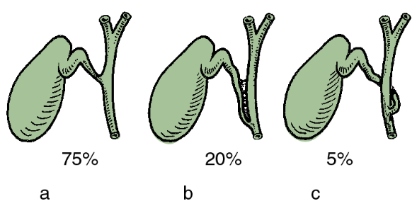
- The mode of union of the cystic duct with the common hepatic duct may be angular, parallel, or spiral
Gallbladder
- Storage organ
- Sit on cystic plate
- Inferior surface of liver
3 parts
- Fundus
- Body
- Neck
- Hartmans pouch caused by gallstones
Adjacent to caudate lobe
Venous
- Drains through cystic plate
Lymphatic
- Cystic node
- Just in front of cystic artery
- Common hepatic
- Coeliac
- Pre-aortic
Innervation
- SNS
- Coeliac ganglia
- Greater, less and least nerves
- Coeliac ganglia
- PNS
- Vagus
Layers
- Mucosa
- Lamina propria
- Thin musclar layer
- Perimuscular connective tissue
- Seroasa - lacks serosal covering along its border with the liver and the perimuscular connective tissue is continuous with the liver connective tissue
- T1a tumours are limited to the lamina propria, which is important as a cholecystectomy can be curative in this subset of patient due to the limited invasion of the gallbladder wall.
Anomilies
- Agenesis 1/8000
- Double
Triangles
- Calot’s Triangle (Original Definition)
- Borders:
- Medial: Common hepatic duct
- Inferior: Cystic duct
- Superior: Cystic artery
- Borders:
- Hepatocystic Triangle
- Borders:
- Medial: Common hepatic duct
- Inferior: Cystic duct
- Superior: Inferior surface of the liver
- Borders:
Further detail
Overview
- Function: Store and concentrate bile
- Capacity: 50mls
Structure
- Fibromuscular sac
- Surprisingly small amount of smooth muscle in its wall histologically
- Mucous membrane:
- Lax areolar tissue
- Simple columnar epithelium
- Arranged in spiral manner in neck and cystic duct (known as the Spiral Valve of Heister)
- Epithelial cells actively absorb water and solutes from bile
- Mucus is secreted by the columnar epithelium – but no goblet cells
- Mucus secreting glands only present in neck
- NB: Gallbladder wall has a submucosa
Three Parts
- Fundus
- Projects beyond lower border of the liver
- Touches parietal peritoneum of anterior abdominal wall at the tip of the 9th costal cartilage where transpyloric plane cross right costal margin at the lateral border of the right rectus sheath
- Lies on the commencement of the transverse colon, just to the left of the hepatic flexure
- Body
- Projects backwards and upwards towards the right end of the porta hepatis
- Is in contact with the 1st part of the duodenum
- Small cystic veins pass from gallbladder into liver substance
- Small bile ducts may also pass from liver into gallbladder
- Neck
- Continues into the cystic duct
- Wall of neck as it joints cystic duct may show a small diverticulum, called Hartmann’s pouch
- Not a feature of a normal gallbladder
- Always associated with pathology, e.g. site of impaction of gallstone
- NB: The neck of the gallbladder is NOT an anterior relation of the Right Kidney
- It actually lies superior and medial to the anterior surface of the Right Kidney
Variations of Gallbladder Anatomy
- Fundus may be folded like a Phyrgian cap (most common congenital abnormality)
- Duplicated with single or double cystic ducts
- Septated with lumen divided into 2 chambers
Relations
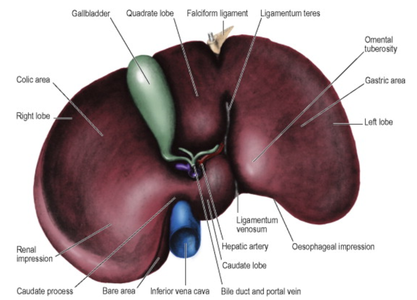
- Lies in gallbladder fossa on visceral surface of right lobe of liver
- NB: The gallbladder bed does not lie entirely in the functional right lobe of the liver because it lies on the division between the right and left lobes
- Adjacent to caudate lobe
Calot’s Triangle
- Boundaries:
- Liver
- Common Hepatic Duct
- Cystic Duct
- Contents:
- Cystic Artery and Vein
- Cystic Lymph Node of Lund
Landmarks during Cholecystectomy
- Rouviere’s Sulcus:
- Fissure visible between Right Lobe (Segment 5) and Caudate Process through which the Right Hepatic Pedicle passes
- Space between the Infundibulum of the Gallbladder and this sulcus is superior to the plane of the CBD and relatively safe for commencing dissection
- Base of Segment 4B (Quadrate Lobe) – Hilar Plate
- Gallbladder rests on Cystic Plate
- Main structures of the Porta Hepatis enter the liver at the Hilar Plate
- Region of Hilar Plate noted by identifying Quadrate Lobe
- Bounded by the Falciform ligament on the Left and the Gallbladder on the Right
Blood Supply Arterial Supply: Cystic Artery
- Branch of Right Hepatic Artery
- Runs across Calot’s Triangle (Liver, Common Hepatic Duct, and Cystic Duct)
- Variations of origin:
- Main trunk of Hepatic Artery
- Left Hepatic Artery
- Gastroduodenal Artery
- Variation in course: pass in front of cystic and bile ducts
Cystic Artery Anatomical Variations
- Cystic Artery arises from the Right Hepatic Artery in Calot’s Triangle and ascends on the left side of the Gallbladder in 70% of cases
- Variations:
- Arises from Common Hepatic Artery, its bifurcation or the Left Hepatic Artery in 20% of cases
- Arises from an Accessory Right Hepatic Artery in 10%
- Arises from Gastroduodenal Artery in < 2.5%
- Significance:
- Right Hepatic Artery passes close to the Gallbladder in 10% of pts with short arterial twigs entering the Gallbladder, rather than a single Cystic Artery
- Artery can thus be damaged when mistaken for the Cystic Artery or when twigs are divided
- Accessory Right Hepatic Artery arises low from the SMA running through Calot’s Triangle posterior and parallel to the Cystic Duct in 8% of pts
- Can be mistaken for a posterior branch of the Cystic Artery
- Right Hepatic Artery passes close to the Gallbladder in 10% of pts with short arterial twigs entering the Gallbladder, rather than a single Cystic Artery
Venous Drainage
- Multiple small veins in gallbladder bed that flow into hepatic veins
- One or more cystic veins may be present (uncommon)
- Run from neck of gallbladder into right branch of Portal Vein
- Cystic veins do not accompany the cystic artery
Lymphatic Drainage
- Drain to nodes in Porta Hepatis, to the cystic node (in Calot’s Triangle at the junction of the cystic and common hepatic ducts), and to a node situated at the anterior boundary of the epiploic foramen
- From these nodes, lymph pass to the coeliac group of preaortic nodes
Cystic Duct
- Length: 2-3cm
- Diameter: 2-3mm
- Runs backwards, downwards, and to the left to join the common hepatic duct
- Cystic duct joins Common Hepatic Duct in front of the right hepatic artery and its cystic branch
- Variations of Cystic Duct insertion to Common Hepatic Duct:
- Runs parallel to and to the right of the Common Hepatic Duct for a variable distance
- Spiral around the Common Hepatic Duct before joining on its left side
- Absent and Gallbladder drains directly into the Common Hepatic Duct
- Common Cystic Duct Variations [2]:
- Right Lateral Insertion
- Anterior Spiral Insertion
- Posterior Spiral Insertion
- Proximal Insertion
- Low Medial Insertion
- Low Lateral Insertion with Common Sheath
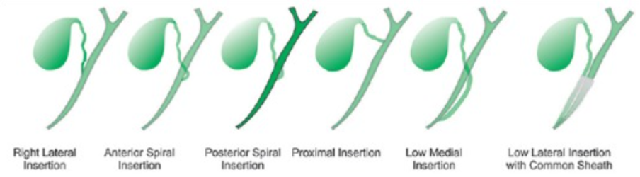
Cystic Duct Variations and Significance
- Three Major Cystic Duct Variants:
- Angular – 75%
- Parallel – 20%
- Spiral – 5%
- Cystic Duct joins to the Right Hepatic Duct or the Right Posterior Sectoral Duct in 2% of cases
- Such ducts particularly at risk during surgery
- Aberrant Hepatic Duct – defined as a duct draining a normal portion of the liver that joints the biliary tree outside the liver
- If Cystic Duct dissected beyond the union with the Aberrant Hepatic Duct or Sectoral Duct, this portion of the liver would not be opacified by the IOC
- Possible to misinterpret IOC as normal
- Only clue would be relative paucity of ducts on the right inferior part of the liver shadow
- If unrecognised, this would lead to excision of the distal end of the RPSD without r ealising it – leading to obstruction or leak
Extra-Hepatic Biliary Tract Overview
- Consists of: Three hepatic ducts (Left, Right, and Common), Gallbladder, Cystic Duct, and Bile Duct
- Right and Left Hepatic Ducts exit liver and join to form the Common Hepatic Duct near the right end of the Porta Hepatis
- Variation: Left Hepatic Duct may run along the base of the quadrate lobe only partially surrounded by liver substance prior to its emergence from the liver
- All extra-hepatic bile ducts are lined by tall columnar epithelium that is mucus-secreting
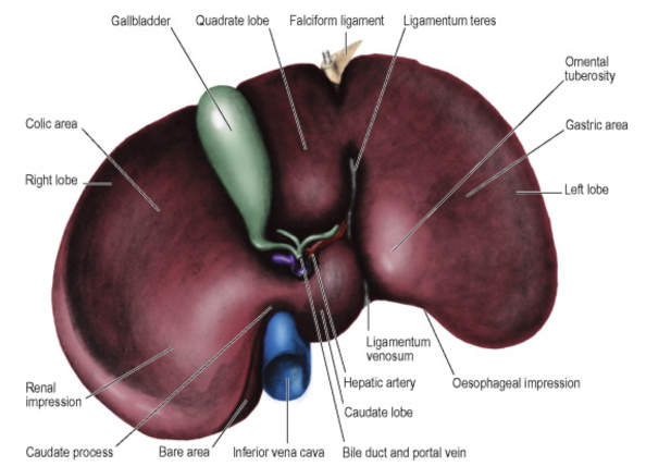
- The Common Hepatic Duct passes down between the two peritoneal layers at the free edge of the Lesser Omentum
- Joined by the Cystic Duct on its right side to form the Bile Duct
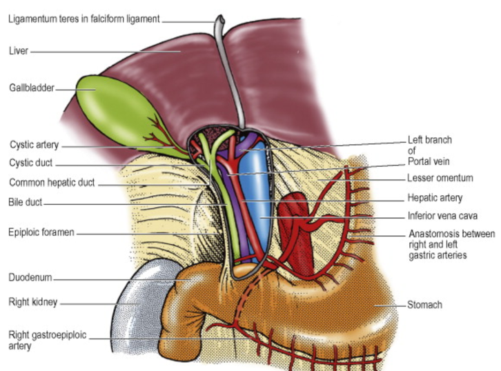
Common Hepatic Duct
- Formed by the Left and Right Hepatic Ducts emerging from the Porta Hepatis and uniting near its right margin
- Joined, usually after about 3cm, by the Cystic Duct to form the Bile Duct
- Junction usually 1-2cm above Duodenum
- Variations:
- Cystic duct runs parallel to and on the right of the Common Hepatic Duct for a variable distance before uniting
- Cystic duct spiralling around the Common Hepatic Duct before uniting on the left side
- Cystic duct absent and gallbladder drains directly into Common Hepatic Duct
- Accessory Right Hepatic Duct which may open into the Common Hepatic Duct, Cystic Duct or Gallbladder
- Right Hepatic Artery usually passes posterior
- Variation: May pass anterior
Bile Duct
- Length: 6-8cm
- Diameter: ≤ 8mm
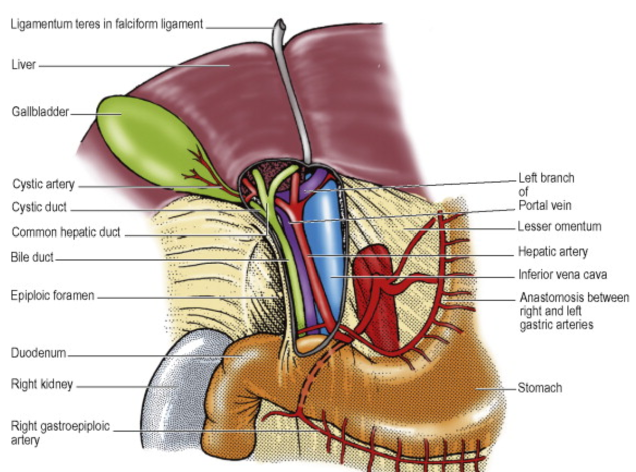
-
Described in 3 parts:
- Upper (Supraduodenal) Third
- Lies in free edge of Lesser Omentum
- Most accessible region for surgery
- Anterior to Portal Vein
- Right of the Hepatic Artery … where the Lesser Omentum forms the anterior boundary of the Epiploic Foramen
- Middle (Retroduodenal) Third
- Posterior to 1st part of Duodenum
- Anterior to IVC
- Slopes down to the right – away from the almost vertical Portal Vein
- Right of the Portal Vein and Gastroduodenal Artery
- Lower (Paraduodenal) Third
- Slopes further to the right
- Lies in a groove in the back of the Head of Pancreas
- Anterior to the Right Renal Vein
- Joins the Pancreatic Duct at about 60° at the Hepatopancreatic Ampulla (of Vater)
- Upper (Supraduodenal) Third
-
Ampulla and ends of the two ducts are each surrounded by smooth muscle – the whole constitutes the Ampullary Sphincter (of Oddi)
-
Variation: Muscle fibres surrounding the ampulla and Pancreatic Duct are absent, leaving only the Bile Duct sphincter
-
Ampulla opens into posteromedial wall of 2nd part of Duodenum at the Major Duodenal Papilla (located 10cm from the Pylorus)
-
Histologically: Tall columnar epithelium, which is mucus-secreting
Blood Supply of the Biliary Tract
- Extrahepatic biliary tract receives small branches from the:
- Cystic Arteries
- Right Hepatic Arteries
- Posterior branch of the Superior Pancreaticoduodenal Artery
- Small veins from the tract drain to the Portal Vein or enter the Liver
Nerve Supply of the Biliary Tract
- Parasympathetic Fibres:
- Mainly from the Hepatic Branch of the Anterior Vagal Trunk
- Stimulate contraction of the Gallbladder
- Thus, Truncal Vagotomy causes hypotonia of the Gallbladder
- Relaxes Ampullary Sphincter
- Sympathetic Fibres:
- From Cell Bodies in the Coeliac Ganglia (with pre-ganglionic cells in the Lateral Horn of Spinal Cord Segments T7-9)
- Inhibits contraction
- Hormonal control of Gallbladder activity (by CCK from neuroendocrine cells of the upper small intestine) is much more important than neural control
- Afferent fibres:
- Includes those for pain
- Mostly run with right-sided sympathetic fibres and reach spinal cord segments T7-9
- Some from the gallbladder may run in the Right Phrenic Nerve (C3-5) through connections between this nerve and the Coeliac Plexus
- Any afferent Vagal fibres are probably concerned with Reflex activities, not pain
- Biliary tract pain usually felt in right hypochondrium and epigastrium, and may radiate to the back in the infrascapular region, in the area of distribution of spinal nerves T7-9
- Phrenic Nerve supply explains the referred pain to the right shoulder