Course
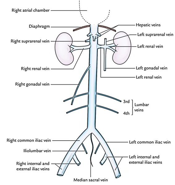
- Begins opposite L5 Vertebra by the confluence of the two Common Iliac Veins behind the Right Common Iliac Artery
- Runs upwards on the right of the Aorta
- Grooves the bare area of the Liver
- Pierces the Central Tendon of the Diaphragm at the level of T8 Vertebra
- Lies on the bodies of the Lumbar Vertebra and the Right Crus of the Diaphragm
- Overlaps the Right Sympathetic Trunk
- Crosses the following structures:
- Right Renal Artery
- Right Suprarenal Artery
- Right Inferior Phrenic Artery
- Partly overlaps the Right Suprarenal Gland and Coeliac Ganglion
-
In the Infracolic Compartment:
- Lies behind the peritoneum of the posterior abdominal wall
- Crossed by:
- Root of the Mesentery
- Right Gonadal Artery
- 3rd Part of the Duodenum
-
In the Supracolic Compartment:
- Lies at first behind the Portal Vein, Head of the Pancreas and the Bile Duct
- Then lies behind the peritoneum that forms the posterior wall of the Epiploic Foramen
-
Above this, lies behind the bare area of the Liver into which it excavates a deep groove
-
Inferior Vena Cava (and its tributaries) do not have valves (EXCEPT the Gonadal, esp. Testicular Veins)
-
Surface Marking: A vertical line 2.5cm to the right of the midline from the Inter-tubercular plane to the 6th costal cartilage
Tributaries Overview
- Tributaries in ascending order above the two Common Iliac Veins are:
- 4th and 3rd Lumbar Veins
- Right Gonadal Vein
- Both Renal Veins
- Right Suprarenal Vein
- Right Inferior Phrenic Veins
- Hepatic Veins
Common Iliac Veins
- Formed in front of the Sacroiliac joint by the union of the Internal Iliac and External Iliac Veins
- External Iliac Veins enter the abdomen on the medial side of its corresponding artery and runs along the pelvic brim
- Left Common Iliac Vein
- Slightly longer than the Right due to the Inferior Vena Cava being located to the right of the midline
- Posteromedial to the Left Common Iliac Artery
- Joins the Right Common Iliac Vein almost at a right angle after bulging forwards across the body of L5 vertebra
- Right Common Iliac Vein
- Almost vertical
- Lies posterior and then lateral to the Right Common Iliac Artery
- Each Common Iliac Vein receives Iliolumbar and sometimes Lateral Sacral veins
- The left usually receives the Median Sacral Vein
- Median Sacral Vein lies on the right side of the Median Sacral Artery
- The left usually receives the Median Sacral Vein
Lumbar Veins
- Accompany the lumbar arteries
- Drain the lateral and posterior abdominal walls
- Anastomotic connections anteriorly with the Epigastric Veins and posteriorly with the Vertebral Venous Plexuses
- The 4th and 3rd Lumbar Veins empty into the Inferior Vena Cava
- The veins from the left pass behind the Aorta
- All expected to lie behind the Sympathetic Trunks
- Occasionally, one or more may lie in front
- The 2nd and 1st Lumbar Veins do not usually reach the Inferior Vena Cava
- They join the Ascending Lumbar Vein which connects the Common Iliac, Iliolumbar, and Lumbar Veins
- Passes vertically upwards behind Psoas and in front of the Lumbar transverse processes
- Each Ascending Lumbar Vein joins the Subcostal Vein to form the Azygos Vein on the right and the Hemiazygos Vein on the left
Gonadal Veins
- Accompany the Gonadal Arteries
- Each is usually paired
- Run up on Psoas with the two venae commitantes uniting
- Right Gonadal Vein:
- Enters the Inferior Vena Cava just below the Renal Vein at an acute angle
- Right Testicular Vein may occasionally join the Renal Vein
- Left Gonadal Vein:
- Invariably joins the Left Renal Vein at a right angle
- Testicular Veins usually have valves at their terminations
- Valves may be present in the Ovarian Veins
Renal Veins
- Lie anterior to the Renal Arteries
- Lie behind the Pancreas
- Joins the Inferior Vena Cava at right angles at the level of L2 Vertebra
- Each emerges from the hilum as 5 or 6 tributaries which soon unite
- Left Renal Vein:
- 3 x as long as the Right Renal Vein (7.5cm vs. 2.5cm)
- Usually crosses anterior to the Aorta
- Receives the Left Suprarenal Vein and Left Gonadal Vein and possibly a Left Inferior Phrenic Vein
- May have to be ligated and divided during surgery for Aortic Aneurysms
- Provided this is done to the right of the point of entry of the Gonadal and Suprarenal Veins, the kidney is not harmed
- Variations:
- Double Left Renal Vein – one vein passing anterior and one posterior to the Aorta
- Only a posterior vein is present
- NB: Left Renal Vein connects the Azygos and Vertebral Venous Systems
- Right Renal Vein:
- Usually only drains its own kidney
- Occasionally is joined by the Right Gonadal Vein
Suprarenal Veins
- Left Suprarenal Vein:
- Runs downwards and medially to the Left Renal Vein
- Right Suprarenal Vein:
- Very short vessel
- Passes horizontally to the posterior aspect of the Inferior Vena Cava behind the Bare Area of the Liver
Inferior Phrenic Veins
- Accompany the arteries on the lower surface of the Diaphragm
- Normally join the Inferior Vena Cava just below the liver
- Left Inferior Phrenic Vein may join the Left Renal or Suprarenal Vein or be double and have different destinations
Hepatic Veins
- Three main Hepatic Veins and several Accessory Hepatic Veins enter the Inferior Vena Cava as it lies in its groove on the back of the Liver
Embryology of the Inferior Vena Cava
- At 4th week gestation – 3 different venous systems form:
- Vitelline system drains the gut
- Umbilical system drains Placenta
- Cardinal system drains the rest of the Embryo
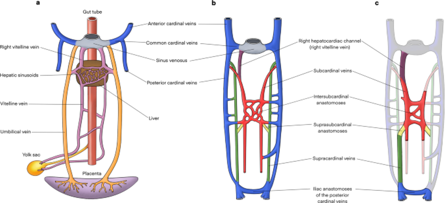
- Infrahepatic IVC develops from a set of 3 paired parallel veins appearing consecutively between 4-8 weeks of life
- Posterior Cardinal Vein
- Subcardinal Vein
- Supracardinal Vein
- Suprahepatic IVC derived from cranial segment of Right Vitelline Vein
- Retrohepatic segment derived from an anastomosis between the cranial segment of the Right Subcardinal Vein and Right Vitelline Vein
- Infra-hepatic/Suprarenal IVC derived from Right Subcardinal Vein
- Renal Collar is formed from anastomoses between the Supracardinal veins posteriorly and the Subcardinal veins anteriorly, with the posterior limb regressing during development
- On the right, the anterior limb is incorporated into the lateral wall of the renal segment of the IVC
- On the left, the anterior limb forms the normal adult Left Renal Vein
- The suprarenal portions of the right and left Supracardinal Veins connect with the Posterior Cardinal veins forming the Azygos and Hemiazygos systems, respectively
- The infrarenal portions disappear on the left but form the infrarenal portion of the IVC on the right
- The Posterior Cardinal Veins mostly regress except for the distal portion, which later become the iliac confluence and future iliac veins, and the proximal portion, which joins the Azygos and the Hemiazygos veins