Overview
- High up on posterior abdominal wall
- At best, only lower poles can be palpated in normal individual
- Oblique lie – long axis parallel to lateral border of Psoas Major
- NB: Long axis slopes downwards and laterally
- Well back in paravertebral gutter such that the Hilum face somewhat forwards as well as medially
- In the erect position, lies opposite the first 3 Lumbar Vertebrae
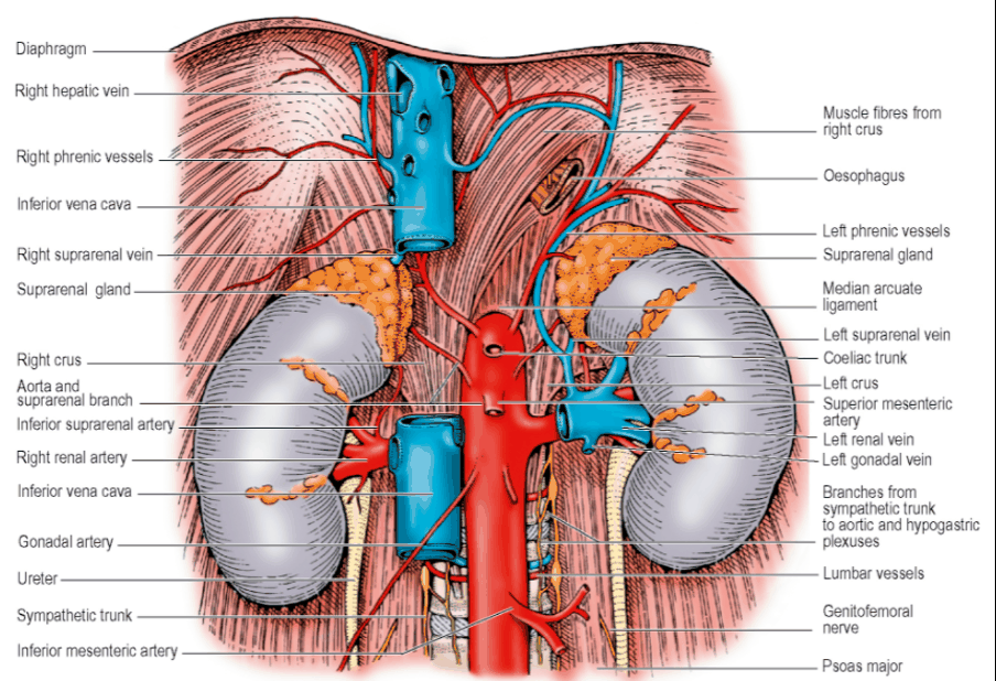
- Normal dimensions: 12 x 6 x 3 cm, Weight 130-150g
- Hilum of Right Kidney just below and Hilum of Left Kidney just above the Transpyloric Plane, 5cm from midline
- Upper pole of Left Kidney overlies 11th Rib
- Upper pole of Right Kidney overlies 12th Rib
- Renal Pelvis emerges from the Hilum behind the vessels to pass down as the Ureter
- Perinephric fat lies outside the Renal Capsule
- Renal Fascia (of Gerota) surrounds the Perinephric Fat
- Condensation of the areolar tissue between the Parietal Peritoneum and Posterior Abdominal Wall
- Restrains the extension of a Perinephric Abscess
- Ascends as a dome over the upper pole of the Kidney and Suprarenal
- Fascial septum separates the two organs
- At the lateral renal border, the anterior and posterior layers fuse
- At the hilum, the fascia is attached to the Renal vessels and the Ureter
- Pus in perinephric space and injections into it do not usually track downwards
- Increasing pressure may force the fascia to rupture and allow such contents to flow downwards retroperitoneally towards the pelvis
Relations
-
Posterior:
- Diaphragm
- Upper poles lies on fibres from the Lateral and Medial Arcuate Ligaments
- Small triangular part of the Costodiaphragmatic Recess of the pleura lies behind the diaphragm
- Quadratus Lumborum
- Psoas Major (overlap medially)
- Transversus Abdominis (overlap laterally)
- Convexity of lateral border of Kidney lies on its aponeurosis origin
- Subcostal Artery and Subcostal Vein
- Subcostal Nerve
- Subcostal vessels and Nerve emerge beneath the Lateral Arcuate Ligament
- Iliohypogastric Nerve
- Ilioinguinal Nerve
- These nerves emerge from the lateral border of the Psoas Major
- Diaphragm
-
Anterior:
- Suprarenal Glands
- Overlap a small part of their anterior surfaces
- Peritoneum of the Hepatorenal Pouch on the right (part of the Greater Sac)
- Peritoneum of the Lesser Sac (Part of the Stomach Bed) on the left medially
- Peritoneum of the Greater Sac on the left laterally (between the Kidney and the Spleen) with the Splenorenal ligament passing forwards between these areas
- Hepatic Flexure on the Right
- Splenic Flexure on the Left
- These colonic segments separate the kidneys from peritoneum
- Coils of Jejunum (with intervening peritoneum)
- Right Colic Artery (ascending branches) – between the peritoneum and kidney
- Left Colic Artery (ascending branches) – between peritoneum and kidney
- Suprarenal Glands
-
NB: Pancreas makes contact with the surface of the Left Kidney
-
Renal Pelvis
- Funnel-shaped commencement of the Ureter
- Normally the most posterior of the three main structures in the Hilum
- Capacity of the average pelvis: < 5mls
Blood Supply and Segments
- Renal Arteries
- Blood flow in excess of 1L per minute
- Leave Abdominal Aorta at right angles
- Lie behind the Pancreas and Renal Veins
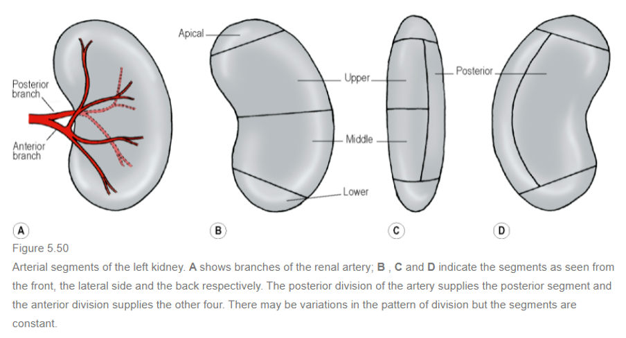
- At the hilum, each artery typically gives an anterior and posterior division
- Renal Segments
- Each kidney has 5 segments
- Posterior division of Renal Artery – supplies posterior segment
- Anterior division of Renal Artery – supplies apical, upper, middle and lower segments
- No collateral circulation between segments
- Abnormal or aberrant renal arteries, e.g. a vessel running from Aorta to the Lower Pole, are actually segmental vessels with an unusual origin (persistence of a foetal vessel)
- Not usually accompanied by veins
- Renal Veins
- Formed from 5 or 6 vessels that unite at the Hilum
- Veins from renal segments communicate with one another (unlike the arteries)
Lymph Drainage
- Drain to Para-Aortic Nodes at the level of origin of the Renal Arteries (L2)
Nerve Supply
- Derived from both parts of the autonomic nervous system
- Sympathetic innervation:
- Sympathetic Preganglionic cells in the spinal cord from T12 to L1 segments
- Send Preganglionic fibres to the thoracic and lumbar Splanchnic Nerves
- Synapse in the Coeliac and Renal Ganglia
- Vasomotor
- Afferent fibres
- Includes those subserving pain
- Accompany sympathetic nerves
- Pathway for pain of renal colic ⇒ Coeliac Plexus ⇒ Splanchnic Nerves ⇒ Sympathetic Trunk (via White Rami Communicantes to T12 to L1 spinal nerves) ⇒ Spinal Cord by the Posterior Nerve Roots
- Possible that some afferents run with vagal fibres which could explain the nausea and vomiting that accompany renal pain
Structure
- Cortex
- Lies beneath the capsule
- Extends towards the Pelvis as the Renal Columns
- Lies between a number of darker and triangular striated areas, the Pyramids of the Medulla
- Apices of several pyramids open into a Renal Papilla
- Each Papilla opens into a Minor Calyx
- Minor Calyces unite to form 2 or 3 Major Calyces which open into the Renal Pelvis
- Functional unit: Nephron
- 1 million in each kidney
- Nephron
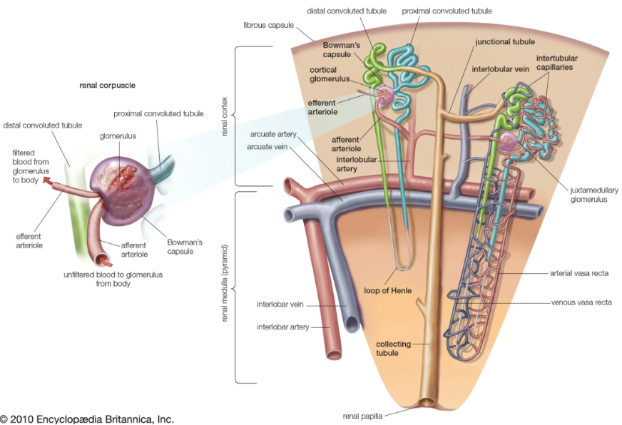
- Consists of a Glomerulus and a Tubule system
- Glomerulus
- Tuft of capillaries
- Supplied by Afferent Arteriole and leaves as an Efferent Arteriole
- Efferent Arteriole breaks up into Peritubular Capillaries surrounding the Proximal and Distal Convoluted Tubules
- Surrounded by very thin epithelial cells (Podocytes)
- Complex projects into Bowman’s Capsule
- Epithelium covering capillaries is continuous with those forming the boundary of Bowman’s capsule which is in turn continuous with the epithelium of the Tubule system
- Epithelium covering capillaries is continuous with those forming the boundary of Bowman’s capsule which is in turn continuous with the epithelium of the Tubule system
-
- Proximal Convoluted Tubule
- Adjacent to Bowman’s Capsule
- Loop of Henle ⇒ Distal Convoluted Tubule ⇒ Collecting Tubule ⇒ Collecting Duct ⇒ Collecting Ducts unite and the largest open at the tip of a Renal Papilla in a Minor Calyx
- Glomeruli and Convoluted Tubules are in the Cortex
- Loops of Henle and Collecting Tubules and Ducts are in the Medulla
- Cells in the Arteriolar system and the Distal Convoluted Tubule constitute the Juxtaglomerular Apparatus – secretes Renin
- Proximal Convoluted Tubule
- Renal Pelvis:
- Transitional epithelium
- Smooth muscle in its wall
- Specialized muscle cells in the walls of the Minor Calyces act as ‘pacemakers’ that initiate contractile waves which pass down the Ureter
Development
-
Three separate excretory organs appear in vertebrate evolution: Pronephros, Mesonephros, and Metanephros
-
Pronephros and Mesonephros – consist of excretory tubules arranged segmentally and empty into the same duct
-
Metanephros – mass of tubules having no segmental arrangement and drains into a new duct that develops specifically for the purpose (the Ureter)
-
Pronephros
- Evanescent (quickly disappearing)
- Duct persists
- Mesonephric Tubules develop and open into the Pronephric Duct a.k.a. Mesonephric (Wolffian) Duct
- Caudal to the Mesonephros, the intermediate cell mass gives rise to a million new tubules forming the Metanephros
-
Metanephros
- Induces a bud, the Ureter, to grow from the caudal end of the Mesonephric Duct
- Ureter bud separates from the Mesonephric Duct
- The Mesonephric Duct forms part of the Bladder, the Vas Deferens, and associated structures
- Ureter bud grows and divides into Calyces of the Pelvis (Major and Minor) and Collecting Tubules of the Medullary Pyramids
- Into these, the Distal Convoluted Tubules of the Mesonephros come to drain
-
Foetal and neonatal kidney have lobulated appearance – reflects the way the Metanephric tissue overlies Tubular budding from the Calyces
-
Definitive Kidney (Metanephros) develops in the Pelvis and is supplied by the Internal Iliac Artery
-
Subsequently migrates to adult position gaining successively new arteries of supply from the Common Iliac and then the Aorta
- Older vessels degenerate as new vessels appear
-
Hilum initially anterior but the kidney rotates 90° medially
Anomalies
- Persistence of foetal lobulation
- No significance
- Persistence of one of the foetal arteries
- Common (30% of individuals)
- Esp. Vessel from the Aorta to the lower pole
- Horseshoe Kidney
- Fusion of the lower poles of the Kidneys
- 1 in 800
- Ureters pass anterior to the isthmus of the kidney substance
- Inferior Mesenteric Artery also passes anterior to isthmus
- Limits ascent of horseshoe
- Polycystic Disease
- 1 in 500
- Both kidneys riddled with cysts
- Hereditary
- May be associated with cysts in the Liver, Pancreas and Lungs
- Renal Agenesis
- Only one kidney
- 1 in 500
- This must be excluded before considering Nephrectomy
Surgical Approach Lumbar Approach - Renal Fascia and Peri-Renal fat incised to expose kidney - Upper pole freed leaving the Suprarenal Gland within its own compartment of the fascia - Overlying peritoneum pushed away forwards and medially - Renal vessels then exposed, ligated and divided - Artery divided before the vein - Ureter transected
- Diseased Right Kidney may adhere to Colon, Duodenum, Inferior Vena Cava or Suprarenal Gland
- Diseased Left Kidney may adhere to the Colon, Spleen, Pancreas or Suprarenal Gland
- Right Renal Vein only 2.5cm long – thus, Inferior Vena Cava very close to the operation area
Percutaneous Renal Biopsy
- Lower pole entered by an approach 2.5cm below the 12th rib
- Needle only advanced when patient is holding their breath so that the kidney is not torn by respiratory movement.
Transplantation
- Donor kidney placed retroperitoneally in the Iliac Fossa with the Hilum parallel to the External Iliac Vessels
- Renal Artery anastomosed to the Internal or External Iliac Artery
- Renal Vein anastomosed to the External Iliac Vein
- Ureter implanted into the bladder