Caecum
Overview
- Projects downwards from the commencement of the Ascending Colon below the Ileocaecal Junction
- Completed covered (usually) by Peritoneum
- Peritoneum reflected downwards to the floor of the right iliac fossa
- Retrocaecal peritoneal space may be shallow or deep
- Often present are 2 Peritoneal Folds from either side of the posterior wall of the Caecum
- Forms the Retrocaecal Recess between them
- The appendix may lie within this recess
- Lies on the Peritoneal floor of the right iliac fossa
- Over the Iliacus and Psoas fasciae
- Over the Femoral Nerve and Lateral Femoral Cutaneous Nerve
- Lower end: At the Pelvic Brim
- When distended: Anterior surface touches Parietal Peritoneum of Anterior Abdominal Wall; When collapsed: Coils of Ileum lie between the two
- Longitudinal muscle is concentrated into 3 flat bands – Taeniae Coli
- One anterior, one posteromedial and one posterolateral
- All 3 converge on the base of the Appendix
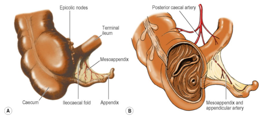
- Internally, the Ileocaecal Junction guarded by the Ileocaecal Valve
- Almost transverse lips may help to prevent reflux into the ileum
- Any possible sphincteric action is poor
Blood Supply
- Branches of Anterior and Posterior Caecal Arteries (from the Ileocolic Artery)
- Posterior Caecal Artery larger
- Gives a branch to the base of the Appendix
- There are corresponding veins
Lymph Drainage
- Nodes associated with the Ileocolic Artery
Appendix
Overview
- Length: Variable – commonly 6 – 9 cm
- Opens into the posteromedial wall of the Caecum 2cm below the Ileocaecal Valve
- McBurney’s Point – one-third of the way up the oblique line that joins the right ASIS to the Umbilicus
- Variable positions:
- Most common position: Retrocaecal
- Second most common position: Pelvic
- Other possibilities: Retrocolic, Retroileal, Paracaecal
- Varying positions change the point of maximal tenderness on clinical examination
- Retrocaecal appendicitis is more likely to present atypically, with less systemic upset, lower likelihood of progression to peritonitis, higher chance of ischaemia, and less or no tenderness in RIF due to overlying gas-filled caecum.
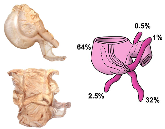
- Three Taeniae of the Caecum merge into a complete longitudinal muscle layer for the Appendix
- Submucosa – contains many lymphoid masses
- Mesoappendix – triangular fold of peritoneum from the left (inferior) layer of the mesentery of the Terminal Ileum
- Bloodless Fold of Treves (a.k.a. Ileocaecal Fold) – a small fold of peritoneum extending from the Terminal Ileum to the front of the Mesoappendix
- Space between the Ileocaecal Fold and the Mesoappendix is the Inferior Ileocaecal Recess
- Vascular Fold of the Caecum – a fold of peritoneum lies in front of the Terminal Ileum, between the base of the mesentery and the anterior wall of the Caecum
- Fold raised up by the contained Anterior Caecal Artery
- Space behind the Vascular Fold of the Caecum is the Superior Ileocaecal Recess
Blood Supply
- Appendicular Artery
- Branch of the Inferior Division of the Ileocolic Artery
- NB: Derived from the Posterior Caecal Artery (Part 1 Bank)
- Runs behind the Terminal Ileum to enter the Mesoappendix
- As it does so, it gives off a recurrent branch that anastomoses with a branch of the Posterior Caecal Artery
- This may replace the Appendicular Artery
- As it does so, it gives off a recurrent branch that anastomoses with a branch of the Posterior Caecal Artery
- Runs first in the free margin of the Mesoappendix and then close to the appendicular wall
- No collateral circulation – i.e. end artery
- Branch of the Inferior Division of the Ileocolic Artery
- There are corresponding veins
Lymph Drainage
- Lymph nodes within Mesoappendix to Ileocolic Lymph Nodes
Ascending Colon
- Length: 15cm
- Extends upwards from Ileocaecal Junction to the right colic (Hepatic) Flexure
- Flexure lies on the inferolateral part of the anterior surface of the Right Kidney
- In contact with the inferior surface of the Liver
- Flexure lies on the inferolateral part of the anterior surface of the Right Kidney
- Lies on Iliac Fascia and anterior layer of the Lumbar Fascia
- Posterior relations:
- Iliacus
- Quadratus Lumborum
- Right Kidney
- Iliohypogastric and Ilioinguinal Nerves
- Front and both sides possess a serous coat
- Runs laterally into the Paracolic Gutter
- Runs medially into the right Infracolic Compartment
- Original embryonic mesentery retained in ~ 10% of adults
- Taeniae Coli:
- Lie in line with those of the Caecum
- Lie anteriorly, posterolaterally, and posteromedially
- If divided between the sacculations, the latter can be drawn part and the bowel wall flattened
- Appendices Epiploicae:
- Small pouches of peritoneum
- Distended with fat
- Blood vessels supplying them from mucosa perforate muscle wall
- Mucous membrane may herniate through these vascular perforations leading to Diverticulosis
Transverse Colon
- Length: 50cm
- Extends from Hepatic to Splenic Flexure
- Splenic Flexure lies at a higher level, under the cover of the left costal margin
- Hangs down anterior to coils of Jejunum and Ileum
- Convexity of Greater Curvature of the Stomach lies in its concavity
- Connected to stomach by the Gastrocolic Omentum
- Greater omentum fused to the Transverse Colon
- Thus, the Greater Omentum appears to hang down from its lower convexity
- Completely invested in Peritoneum
- Hangs free on the Transverse Mesocolon
- Attached from the inferior pole of the Right Kidney ⇒ Across the 2nd Part of the Duodenum ⇒ Pancreas ⇒ Inferior Pole of Left Kidney
- Taeniae coli - due to some rotation of the gut wall at the flexures, the anterior taenia of ascending and descending colons lie posteriorly while the other two lie anteriorly, above and below
- Appendices Epiploicae – larger and more numerous than the Ascending Colon
Descending Colon
- Length: 25cm
- Extends from Splenic Flexure to the Pelvic Brim (5cm above the Inguinal Ligament)
- Plastered to the posterior abdominal wall throughout its course by peritoneum
- Mesentery still present in 20% of adults
- Lies on the Lumbar Fascia and Iliac Fascia
- Splenic Flexure:
- Lies on the lateral surface of the Left Kidney
- Lies below and in contact with the Tail of the Pancreas and the Spleen
- Phrenicocolic Ligament:
- A fold of peritoneum
- Attaches the Splenic Flexure to the Diaphragm at the level of the 11th and 12th ribs
- Requires division during surgical mobilization
- Spleen needs to be safeguarded as it lies in contact with the upper surface of this ligament
- Mobilization of the Descending Colon carried out by dividing the peritoneum along the white line of Toldt
- Taeniae Coli: Lie one anterior and two posterior (medial and lateral)
- Appendices Epiploicae numerous and diverticulosis common
Sigmoid Colon
- Length: 40cm
- Extends from the Descending Colon at the Pelvic Brim to the commencement of the Rectum in front of the 3rd piece of the Sacrum
- Completely invested in Peritoneum
- Hangs free on the Sigmoid Mesocolon
- Lies, usually, in the pelvic cavity
- Coiled in front of the Rectum
- Lying on the Peritoneal surface of the Bladder and Uterus
- Congenital peritoneal adhesions frequently found between lateral aspect of the Pelvic Mesocolon and the Parietal Peritoneum of the Left Iliac Fossa floor – requires division during surgical mobilization of the Sigmoid Colon
- Taeniae Coli
- Wider than elsewhere
- Meet to clothe the terminal part of the Sigmoid Colon in a complete longitudinal coat
- Well-developed Appendices Epiploicae
- Diverticulosis – most common in his part
Blood Supply of Colon
-
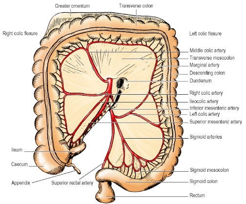
Ascending Colon and Proximal 2/3rds of Transverse Colon:
- Ileocolic, Right Colic, and Middle Colic Branches of the Superior Mesenteric Artery
-
Remainder of the Colon:
- Left Colic and Sigmoid Branches of the Inferior Mesenteric Artery
-
Marginal Artery (of Drummond):
- Formed by the anastomotic branches near the inner margin of the whole colon
- Form short vessels that run into the gut wall
- Weakest link in the chain – near the Left Colic Flexure, i.e. between the Middle and Left Colic Branches (i.e. between midgut and hindgut vessels)
- Inner Arterial Arc (Of Riolan) between the Ascending branch of the Left Colic Artery and the trunk of the Middle Colic Artery may supplement the blood supply in this region
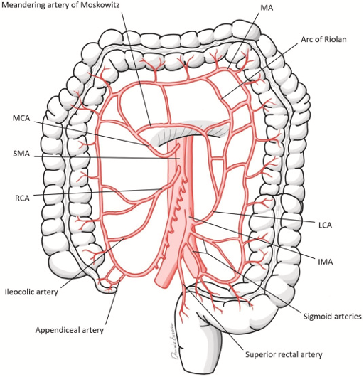
- Inner Arterial Arc (Of Riolan) between the Ascending branch of the Left Colic Artery and the trunk of the Middle Colic Artery may supplement the blood supply in this region
-
Veins correspond to Arteries ⇒ Superior or Inferior Mesenteric Veins ⇒ Portal Vein
-
Some anastomosis between Portal and Systemic Venous drainage where the Ascending and Descending Colon are in contact with the posterior abdominal wall
Lymph Drainage
- Follow arteries
- Drain to Superior or Inferior Mesenteric Nodes
Nerve Supply
- Parasympathetic supply:
- Up to near the Splenic Flexure: From the Vagi
- From the Hindgut (Splenic Flexure onwards): Pelvic Splanchnic Nerves
- As for Rectum, pain fibres from the Descending and Sigmoid Colon run with the Parasympathetic fibres
- Sympathetic supply: Spinal cord segments T10-L2
- Vasoconstrictor fibres
- Pain fibres accompany the above
- Peri-umbilical pain if from the midgut derivatives
- Hypogastric pain if from the hindgut
Colectomy Anatomy
- Right Hemicolectomy:
- From Terminal Ileum to the proximal part of the Transverse Colon
- Ligation of the Ileocolic and Right Colic arteries adjacent to their Superior Mesenteric parent
- Transverse Colectomy:
- From the Right Colic Flexure to the Left Colic Flexure including the Transverse Mesocolon and Greater Omentum
- Ligation of the Middle Colic Vessels
- Left Hemicolectomy:
- Left end of the Transverse Colon to part of the Sigmoid Colon
- Ligation of the Left Colic and Upper Sigmoid Vessels
- Sigmoid Colectomy:
- From the lower Descending Colon to the Rectum
- Ligation of the Lower Left Colic and Sigmoid Vessels
Rectum
Overview
- Length: 12cm
- Continuous with the Sigmoid Colon at the level of the 3rd piece of Sacrum
- Transition is gradual
- In this region, Sigmoid Mesocolon ends and Rectum has no free Mesentery
- Taeniae of Sigmoid Colon gradually broaden to form wide anterior and posterior muscular bands which meet laterally to give the Rectum a completely outer layer of longitudinal muscle (Rectum has no sacculations)
- No Appendices Epiploicae in the Rectum
- Follows the posterior concavity of the Sacrum
- Three lateral curves/flexures
- Upper and lower curves convex to the right
- Middle curve convex to the left
- Thus, middle part appears to bulge to the left
- NB: Has an ampulla that is concave to the left
- Three sickle-shaped transverse Rectal Folds (a.k.a. Rectal Valves of Houston) correspond to these curves
- Incorporate the circular muscle of the wall
- Are not confined merely to the mucous membrane (like the circular folds of the Duodenum or Jejunum)
- Middle fold (largest) projects into the lumen from the right wall of the Rectum to form the floor of the Rectovesical or Rectouterine Pouch
- About 8cm from the anal orifice
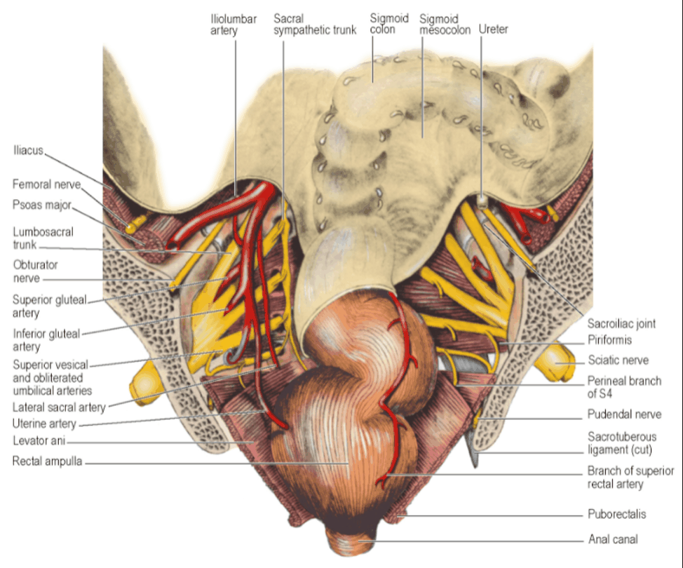
- Rectal Ampulla – lowest part that is slightly dilated (Concave to the Left)
- Rectum turns downwards and backwards as the Anal Canal 2-3cm in front of the tip of the Coccyx
- Anorectal Junction slung forwards by U-loop of the Puborectalis
- Puborectalis merges with the top of the External Anal Sphincter
- Forms a palpable ledge (Anorectal ring) on rectal exam
- Puborectalis merges with the top of the External Anal Sphincter
Relations Anterior Relations in Male
- Rectovesical Pouch – occupied by Small Intestine or Sigmoid Colon
- Bladder
- Seminal Vesicles
- Prostate
- Ends of each Ureter
- Ends of Vas Deferens
Anterior Relations in Female
- Rectovaginal pouch – occupied by Small Intestine or Sigmoid Colon
- Vagina
- Cervix – Projecting into the Fornix of the Vagina
Posterior Relations
- The following structures separate the distended Rectum from the Sacrum and Coccyx
- Ganglion Impar
- Sacral part of the Sympathetic Trunk
- Piriformis Muscle NB: NOT the Inferior Hypogastric Plexus
Mesorectum
- Connective tissue and fat around the Rectum
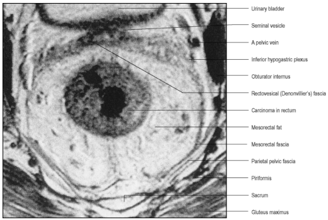
- Mesorectal fascia: Visceral fascia surrounding this tissue
- Bulkier posteriorly – where it tends to be grooved in the midline
- Contents:
- Superior Rectal Artery and branches
- Superior Rectal Vein and its tributaries
- Lymphatic Vessels
- Lymph Nodes
- Relatively avascular plane exists between Mesorectal Fascia and Parietal Pelvic Fascia
- This is the plane of surgical dissection in TME surgery
- Plane most evident posteriorly
- Plane minimal laterally where the Inferior Hypogastric Plexus lies tangentially on the surface of the Mesorectal Fascia
- Crossing this interface are autonomic nerve fibres from the plexus to the Rectum and occasional small Middle Rectal Vessels
- Surgical definition of surrounding connective tissue from the Mesorectum comprises the iatrogenic ‘Lateral Ligament’ of the Rectum – this is not seen on MRI or CT scanning
- Oncological resection involves carefully removing the entire mesorectum contained within the Mesorectal Fascia in order to ensure all lymphatic tissue is removed for both prognostic staging and therapeutic purposes
- Waldeyer’s Fascia (a.k.a. Rectosacral Fascia) originates from the Presacral Parietal Fascia at the S2 to S4 level fusing with the Rectal Visceral Fascia at the posterior aspect of the Rectum
- Divides the Retrorectal space into superior and inferior compartments
- Suspends the Rectum in the hollow of the Sacrum
Peritoneum Coverage of Rectum
- Covers upper 1/3 of the Rectum at front and sides
- Covers middle 1/3 only at the front
- Lower 1/3 is below the level of the Peritoneum
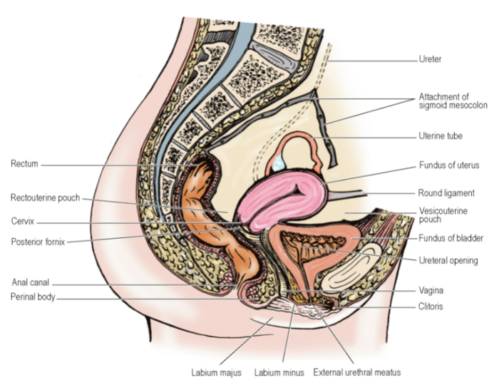
- Peritoneum reflected forwards onto the upper part of the Bladder (in males) or upper Vagina to form the Rectovesical Pouch or Rectouterine Pouch (of Douglas).
- These pouches form the lowest parts of the Peritoneal Cavity
- Located 7.5 and 5.5cm from the Anal margins in male and females, respectively
- Within reach of the finger tip on rectal exam
- Normally occupied by coils of Small Intestine or Sigmoid Colon
Rectovesical Pouch
- Anterior:
- Uppermost part of the Base of the Bladder
- Tops of Seminal Vesicles
- Inferior:
- Rest of the Bladder Base
- Seminal Vesicles
- Prostate
- Ends of each Ureter
- Ends of Vas Deferens
Rectogenital Septum a.k.a. Rectovesical Fascia of Denonvilliers
- Condensation of fascia between these structures and the Rectum
- Connected to the floor of the Rectovesical Pouch above and the Apex of the Prostate below
- Fusion of the anterior and posterior walls of the pouch may account for the origin of this septum
- Distinct whitish appearance in the living
- Closer to the Rectum than to the Seminal Vesicles and Prostate
- Usually removed in rectal excision surgery
Rectouterine Pouch
- Anterior:
- Uppermost part of the Vagina (Fornix, with the Cervix projecting into it)
- Below the peritoneal reflection:
- More of the Vagina, with the Rectogenital Septum intervening
- Thin Rectovaginal/Rectogenital septum fuses with the Perineal Body below
Rectourethralis Muscle
- Slips from the longitudinal Rectal Ampulla
- Pass forwards to the Perineal Body and Sphincter Urethrae in male
- Must be cut at operations for excising Rectum and Anal Canal
Blood Supply
-
Superior Rectal Artery
- Lower end of Inferior Mesenteric Artery enters Sigmoid Mesocolon
- Changes name to Superior Rectal Artery on crossing the Pelvic Brim
- Crosses the Left Common Iliac Vessels medial to the Ureter NB: This does NOT occur at the Sacroiliac Joint
- Descends in the base of the medial limb of the Mesocolon
- Divides into two branches at the level of S3 Vertebra (where the Rectum begins) that descend on each side of the Rectum and subdivide into smaller branches
- Vessels pierce muscular wall and supply the whole thickness of Rectal Wall including mucous membrane
- Continue in the Submucosa into the Anal Canal
- Anastomose with branches of the Inferior Rectal Artery
-
Middle Rectal Artery
- Present in only one in five people
- Small
- Supply only the muscle of the mid and lower Rectum
-
Inferior Rectal Artery
- Capable of supplying the Rectum from below to a level at least as high as the Peritoneal reflection from its anterior surface
-
Median Sacral Vessels
- May make an unimportant contribution to the posterior wall in the region of the Anorectal Junction
- Main clinical significance: May cause bleeding at operations in this region
-
Veins correspond to arteries
- Main route of venous drainage: Via Superior Rectal Vein to the Inferior Mesenteric Vein – crosses Pelvic Brim between the Inferior Mesenteric Artery and Ureter
- Inferior Rectal Veins drain to the Internal Pudendal Veins
- Anastomose freely with one another
- Forms an Internal Rectal Plexus in the Submucosa
- Lower end of Internal Plexus continuous with vascular cushions of the Anal Canal
- Forms an External Rectal Plexus outside the muscular wall
Lymph Drainage
- Mainly upwards
- Lymphoid follicles in mucous membrane ⇒ Epicolic Nodes on Rectum surface ⇒ Pararectal Nodes in Mesorectum ⇒ Pre-aortic Nodes (via nodes along the Inferior Mesenteric Artery)
- Lymph drainage from lower Rectum to Internal Iliac Nodes along Middle Rectal and Inferior Rectal Arteries and along Median Sacral Artery: minimal – unlikely to be a route for metastatic spread of cancer that has not breached the mesorectal fascia
Nerve Supply
-
Sympathetic supply:
- Fibres that accompany the Inferior Mesenteric and Superior Rectal Arteries from the Inferior Mesenteric Plexus
-
Parasympathetic supply:
- S2, S3 and S4 and by Pelvic Splanchnic Nerves via the Inferior Hypogastric Plexus
- Motor to the Rectal muscle
- Afferent fibres convey sensation of distension
-
Pain fibres – accompany both Sympathetic and Parasympathetic supplies
-
Nervi Erigentes, a.k.a. Pelvic Splanchnic Nerves
-
Pre-ganglionic parasympathetic nerve fibres
-
The Splanchnic Nerves come into close proximity on the lateral surfaces of the Mesorectal Fascia, which are excised during APR surgery and are at risk of being damaged.
Rectal Exam
- Palpable structures in both genders:
- Coccyx and Sacrum posteriorly
- Ischial spines laterally
- Anorectal ring posteriorly – shelf-like projection over the tip of the finger that can be hooked when the patient bears down
- Males:
- Prostate felt anteriorly
- Normal Seminal Vesicles are not usually palpable
- Females:
- Cervix felt through Vaginal wall
- Uterosacral ligaments laterally
- Ovaries (sometimes)
Development
- Rectum and Anal Canal derived from Anorectal Canal (Dorsal part of the Cloaca) and the Proctodeum
- Anal membrane breaks down at a site probably represented by the Pectinate Line in the Anal Canal
- Anal valves indicate remains of the membrane
- Part of Anal Canal continuous with Rectum above Pectinate Line is Endodermal
- Part of Anal Canal below Pectinate Line derived from Proctodeum and Ectodermal
Surgical Approach
-
For surgical management of rectal carcinoma:
-
Anterior Resection or Total Mesorectal Excision (TME)
- Access through anterior abdominal wall
- Most or all of Rectum and surrounding Mesorectum removed
- Usually with Sigmoid Colon and Mesocolon
- Lower part and/or anal canal left behind to anastomose with the mobilized Descending Colon
- Rectovesical (Rectovaginal) Fascia usually removed with the rectum after division of its attachment to the prostate/perineal body
- Rectosacral Fascia divided
- Superior Hypogastric Plexus and both Inferior Hypogastric Plexuses kept intact to safeguard sexual function and urinary voiding
- Inferior Mesenteric Artery divided close to its Aortic origin to ensure removal of all lymph nodes along its course and preserving the Inferior Mesenteric Plexus on the Aortic surface
-
Abdominoperineal Resection (APR)
- Complete excision of Rectum and Anal Canal
- Includes dividing the Pelvic Floor (Levator Ani)
- Ischioanal Fossae entered via elliptical incisions either side of the Anus
- Coccyx dislocated or excised
- Rectosacral Fascia divided from below
- Anteriorly, dissection extends up to Transverse Perineal Muscles and Perineal Body
- Rectourethralis muscle and Rectogenital Septum divided
- Plane between the septum and Prostate or Vagina entered from below
- Cylinder of tissue removed through the Perineum
- Pelvic floor repaired by transferring a flap of muscle, e.g. from Gluteus Maximus or Rectus Abdominis
- Terminal Colostomy made in left iliac fossa