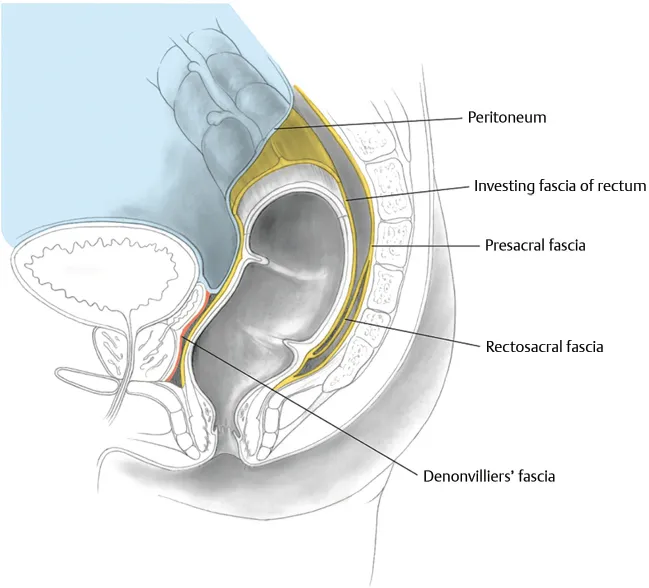
The rectum is the part of the gut between the sigmoid colon and the anus. It is usually 12cm in length and has three characteristic folds, forming valves (of Houston) on the inside; two to the left and one to the right. It commences where the teniae of the sigmoid converge to form a full muscular coat, usually at the level of S3. It is also where the colonic mesentery ends; the anterior rectum is covered by peritoneum though the posterior and lateral wall reside within the mesorectum. Anteriorly, the peritoneum forms a cul-de-sac. The rectum terminates where puborectalis slings it forwards, from this point the gut is the anal canal.
Blood supply is via the superior, middle, and inferior rectal arteries, from the IMA, internal iliac, and pudendal arteries respectively. Venous drainage is portal above and systemic below, and shunts may occur here. Nerve supply is via the pelvis splanchnics for parasympathetic supply and the inferior hypogastric plexus for sympathetic supply. Lymphatic drainage is to the IMA nodes.
The upper rectum is intraperioneal and the middle rectum is intraperioneal only anterioly.
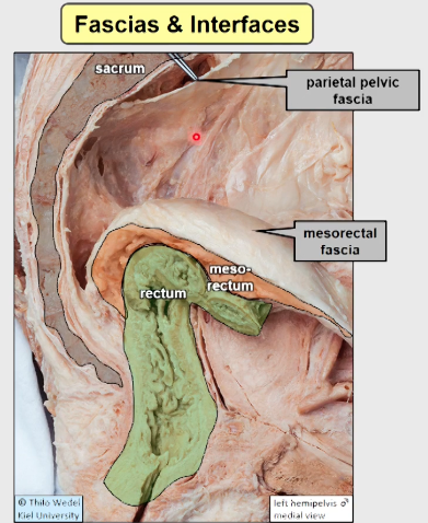
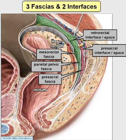
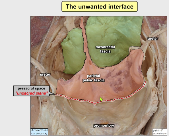
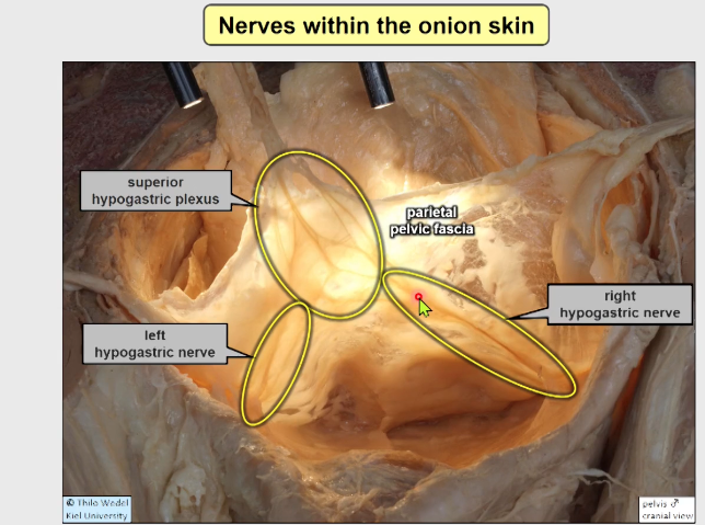
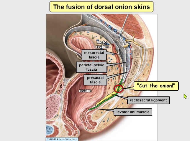
Three layers of fascia posteriorly
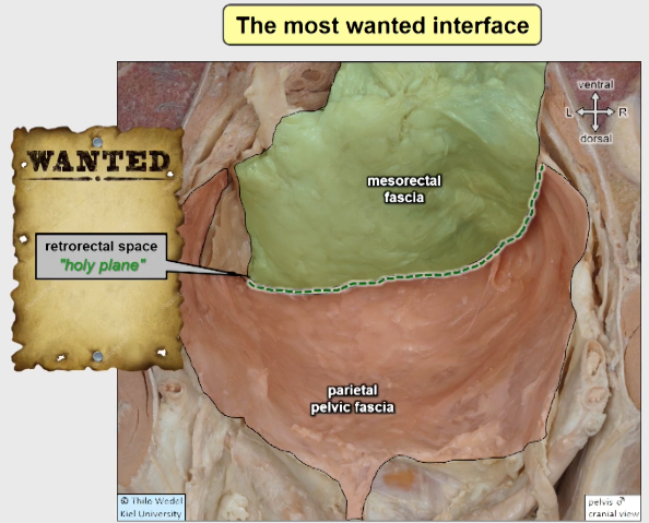
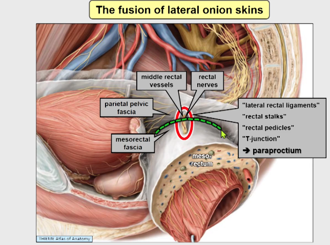
- Mesorectal fascia (visceral pelvic fascia or fascia propria of the rectum) - FPR
- Parietal pelvic fascia (pre hypogastric fascia) - PHF
- Pre sacral fascia
Waldeyers fascia - rectosacral fascia - RSF
- This is fascial layer which is the end of the parietal pelvic fascia which attaches to the mesorectal fascia at the level of S4
- This divides the retrorectal space in to a presacral (cranial) and precoccygeal (caudal) space i.e upper and lower portion
TME - aim to dissect between the mesorectal fascia and the parietal pelvic fascia
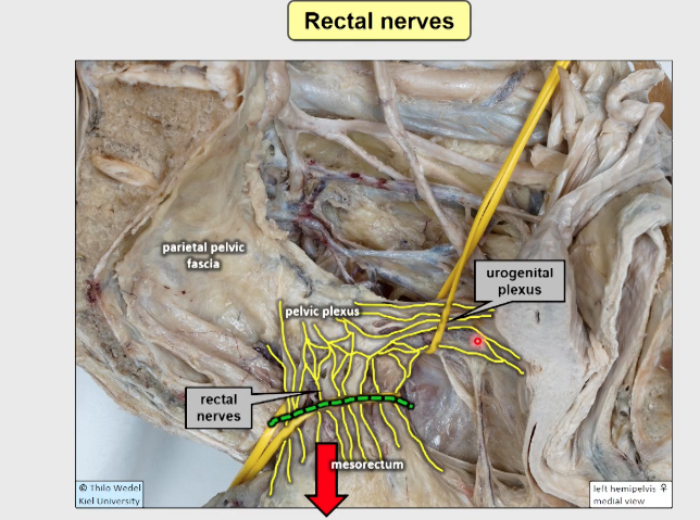
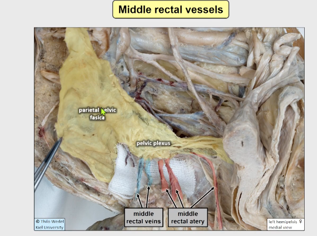
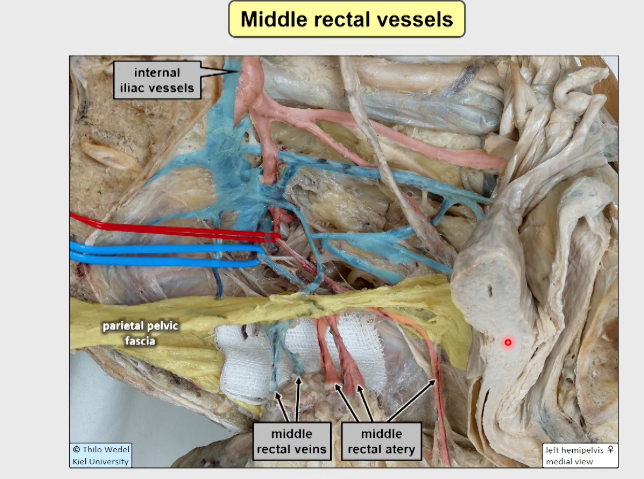 Pelvic side wall
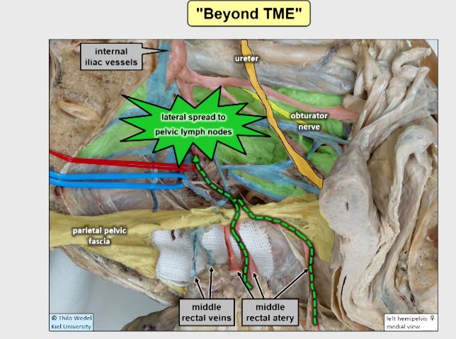
Anterior
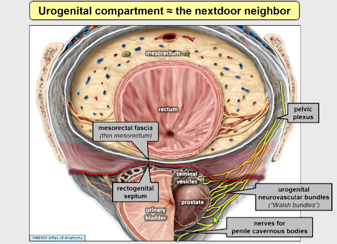
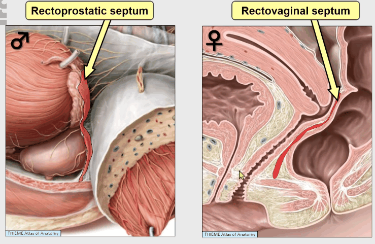
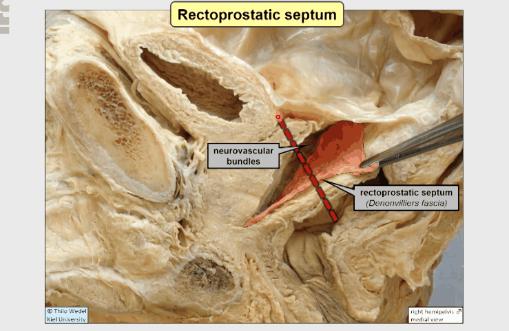
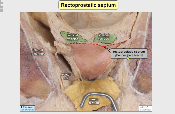
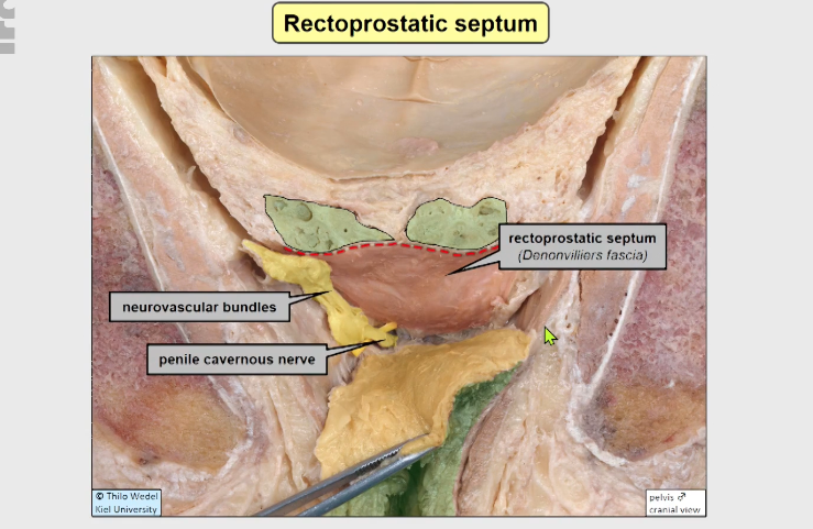
Source: Video
https://academic.oup.com/gastro/article/doi/10.1093/gastro/goac001/6524804