Section: Skin and soft tissue Sub-section: Skin infections and breakdown Curriculum: Curriculum, page 54
Causes of hand infections
- Trauma
- Animal bites
- Human bites.
- Contaminated lacerations
Risk factors
- Diabetes
- Smokers
- Immunosuppression
- Intraveous drug use.
General
- Swelling of dorsum is common with palmar infections
- Lymphangitis of the upper limb in association with hand infection = rapid spread
- USS can be used
- Always immobilise & elevate
- Drainage of wounds parallel to neurovascular structures
- Zig-zag wounds across creases
- Avoids contractures
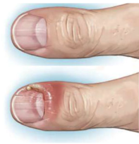
Paronychia
- Infection of soft tissue folds surrounding lateral aspects of fingernail
- Usually from over vigorous grooming
- Early infection
- Treat with splinting and oral antibiotics (Augmentin)
- Advanced infection
- Drain under digital block by lifting infected fold off nail with a Scalpel
Felon
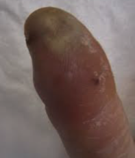
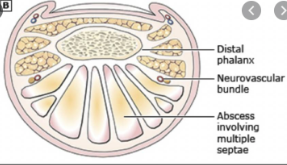
- Painful infection of volar fingertip,
- Usually caused by penetrating injury
- (get x-ray to look for foreign body).
- Infection usually contained by fibrous septa joining periosteum of distal phalanx to dermis.
- Infection can track down septa to cause osteomyelitis
- Early felons treat with splinting, elevation, antibiotics (Augmentin)
- Advanced infection - treat - drain under digital block, via volar longitudinal incision
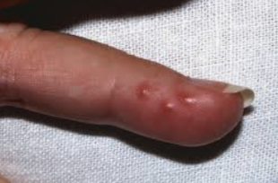
Herpetic Whitlow
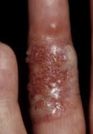
- Infection by HSV 1 and 2, due to contact with infected body fluids
- Dental & healthcare workers
- Clinical
- Fever & malaise, then burning, pain, or tingling of digit,
- Clear vesicles after 7 to 10 days which coalesce then rupture and ulcerate.
- Self-limiting. Recurs in 50%
- Confirm diagnosis by
- Culturing the virus from vesicular fluid,
- Assessing immunofluorescent serum antibody titres or performing a Tzanck smear
- (none of these usually required)
- Management if overly symptomatic
- Can de-roof blisters for pain.
- Topical or oral acyclovir to shorten symptoms and prevent recurrence
Suppurative Flexor Tenosynovitis
- Infection of flexor tendon sheath
- Between A1 pulley at metacarpal head and the insertion of FDP at distal phalanx.
- Infection of thumb and little finger sheath can communicate proximally with radial and ulnar bursae.
- Usually Staph and Streptococcus.
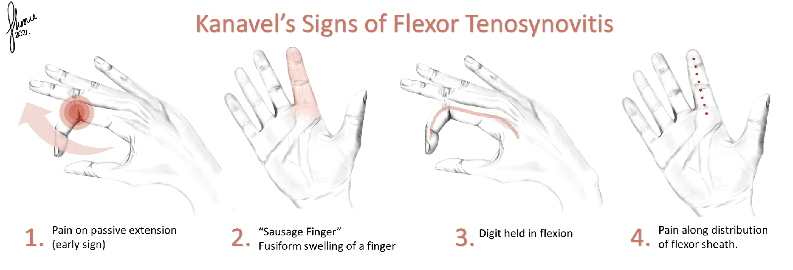
- Kanavel’s 4 cardinal signs
- Symmetrical enlargement of finger
- Semi-flexed position (allows synovial sheath its maximal volume & eases pain)
- Pain along flexor tendon sheath (in palm too)
- Severe pain on passive extension
- Early infection - treat with antibiotics. (Augmentin)
- Advanced infection treat with incision (short sagittal mid-line) and drainage and continuous irrigation (antibiotic solution) with catheter in synovial bursa and have an exit incision in the palm, or more extensive exposure of entire tendon sheath
Deep Hand Infections
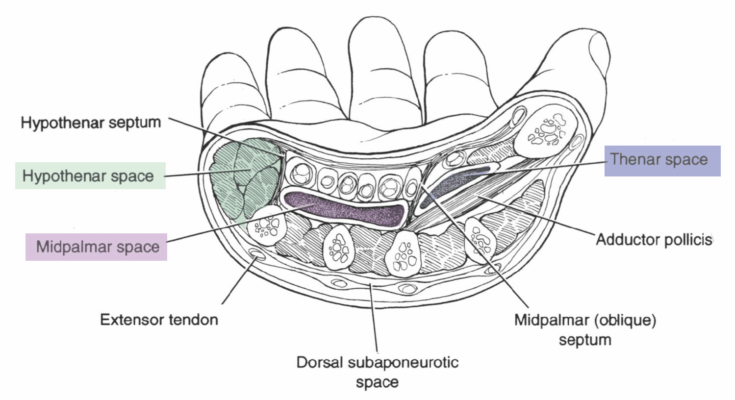
- Infection in one of six sub-fascial spaces:
- Palmar aponeurosis divides the compartments
- Thenar
- Hypothenar
- Mid-palmar
- 3x web space
- Paronaʼs (volar forearm)
- Dorsal sub-aponeurotic
- Palmar aponeurosis divides the compartments
- Usually due to penetrating injury
- Staph or Streptococcus.
- (Augmentin)
- (Augmentin)
Thenar Space
- Anatomy
- Overlies adductor pollicis
- Ulnar boundary is the mid palmar septum (between palmar aponeurosis and third metacarpal)
- Radial boundary is where adductor pollicis joints onto the first metacarpal
- Anterior to the thenar space is the flexor tendon of index finger.
- Infection can spread dorsally through adductor pollicis
- Clinical
- Pain on moving thumb.
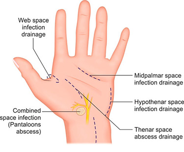
- Drain
- Can drain from dorsal aspect of volar aspect
- Incision 1cm proximal to the web space, extending proximally
- Blunt dissection continues to adductor pollicis and the abscess is drained.
- You may encounter the motor branch of the median nerve.
Hypothenar Space
- Anatomy
- Is located between the hypothenar fascia and 5th metacarpal.
- It contains the hypothenar muscles within it
- It ulnar border is the flexor retinaculum.
- Is radial border is the hypothenar septum (septum between 5th metacarpal and palmar aponeurosis)
- Drain
- Along the line of the 5th metacarpal
Mid-Palmar Space
- Anatomy
- Is a continuation of the carpal tunnel.
- Deep to the palmar aponeurosis and flexor tendons (within their sheaths)
- Superficial to the palmer interossei.
- Ulnar aspect is the hypothenar septum (septum between 5th metacarpal and palmar aponeurosis)
- Radial aspect is the mid palmar septum (between the palmar aponeurosis and 3rd metacarpal)
- Drainage of mid-palmar space abscess
- Approached palmar aspect.
- Transverse incision and mid-palmer crease
- Blunt dissection either side of the flexor tendons until abscess is drained.
Dorsal Sub-Aponeurotic Space
- Anatomy
- Lies dorsal to the extensor tendons of the hands.
- Drainage of a dorsal hand space abscess
- Most dorsal hand abscesses can be drained in the ED under a local field or regional block.
- Longitudinal incision should be biased toward the region over the second metacarpal space of the space between the 4th and 5th metacarpal to avoid the extensor tendons.
Web Space
- Drain via volar zigzag ± dorsal incision, dividing transverse carpal ligament
- There are three web spaces
- One between the bases of the proximal phalanges
Parona’s Space
- Deep to flexor tendons of forearm
- Communicates with radial and ulnar bursa and mid-palmar space
Chronic Infections
- Paronychia – Candida
- (mycobacterium or gram-negative)
- May need to marsupilise to clear
- Tenosynovitis mycobacterium or fungi
- May need to excise involved synovium
- Need to differentiate from chronic granulomatous synovitis
- Sarcoidosis, amyloidosis, gout, RA