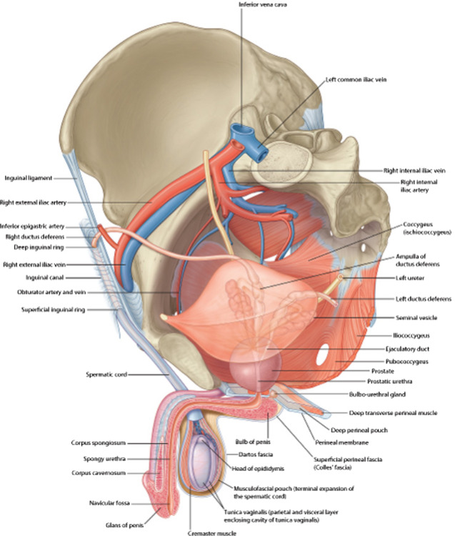
Layers
- There are 7 layers (from outside to inside)
- Skin
- Dartos Muscle
- External Spermatic Fascia
- Derived from the aponeurosis of the external oblique muscle
- Cremaster Muscle and Fascia
- Derived from the internal oblique muscle
- Internal Spermatic Fascia
- Derived from the transversalis fascia
- Tunica Vaginalis
- A double-layered serous membrane derived from the peritoneum.
- Tunica Albuginea (of the Testes)
- The dense, fibrous outer covering of the testes itself
Structures
- Epididymis
- Attached to posterolateral surface
- Vas deferens
- Arise from lower pole of epididymis and runs up medial to it behind the testis
- Appendix Testis – minute sessile cyst attached to the upper pole of the testis
- Remnant of the paramesonephric duct
- Structure
- Front and sides of testis lie free in serous space formed by the overlying serous membrane, the Tunica Vaginalis (a remnant of the fetal Processus Vaginalis)
- Tunica Vaginalis also covers the anterolateral part of the Epididymis and lines the Sinus of the Epididymis, a slit-like space between the testis and Epididymis
- Testis + Epididymis + Tunica Vaginalis lie within thin membranes which are downward prolongations of the coverings of the spermatic cord.
- Median Scrotal Septum separates right and left sides
- Average dimensions: 5cm length, 2.5cm breadth, 3cm AP
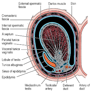
Coverings of the Spermatic Cord/Testicle
- Tunica vaginalis
- Actually runs in cord and bliterated
- Deepest layer surrounding testicle
- Derived from peritoneum
- Internal Spermatic Fascia
- Derived from transversalis fascia at the deep ring
- Cremasteric fascia (and muscle)
- Derived from internal oblique
- Actually spiral around cord, to testicle, and back up
- Accounts for cremasteric reflex
- External spermatic fascia
- Derived from External Oblique Aponeurosis
- Picked up at crura of superficial ring
- Not part of the cord in the inguinal canal
Blood Supply
- Testicular Artery
- Origin: Aorta (at L2 level)
- Course: Runs in spermatic cord, gives off a branch to the Epididymis, reaches back of testis, divides into medial and lateral branches
- In region of Epididymis, there is an anastomosis between the Testicular, Cremasteric and Ductal arteries
- If main artery divided, smaller vessels may not completely sustain the testis and atrophy may occur
- Ischaemic necrosis unlikely
- Venules reach the Mediastinum Testis
- Pampiniform Plexus
- Mass of intercommunicating veins passing upwards in the spermatic cord formed from the venules
- In inguinal canal – plexus separates out into ~4 veins which join to form 2 that leave the Deep Inguinal Ring, becoming a single vein on Psoas Major on the posterior abdo wall
- Left Testicular Vein – joins the Left Renal Vein at a right angle
- Right Testicular Vein – drains directly into the IVC at an acute angle
- Testicular veins usually have valves
- Varicocoele (varicosities of the Pampiniform and Cremasteric Veins) occurs much more frequently on the left than the right
- Mass of intercommunicating veins passing upwards in the spermatic cord formed from the venules
- Cremasteric artery
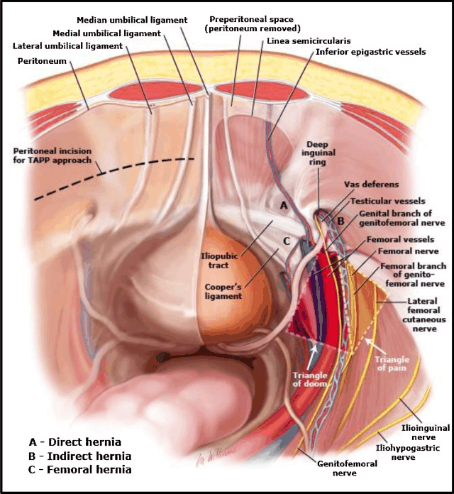
- originates from the inferior epigastric artery
Lymphatic Drainage
- Travel back with the Testicular Artery to Para-aortic nodes alongside the Aorta at the level of the origin of the Testicular Arteries (L2 vertebra, i.e. just above the umbilicus)
- Testicular lymph, thus, does not drain to inguinal nodes
- Overlying Scrotal Skin does
Nerve Supply
- Sympathetic nerves – most connector cells lie in T10 segment of spinal cord
- Pass mainly in the Lesser Splanchnic Nerve to the Coeliac Ganglia and the efferent fibres synapse there
- Post-ganglionic grey fibres reach the Testis along the Testicular Artery
- Sensory fibres – share same sympathetic pathway – run up along Testicular Artery and through the Coeliac Plexus and Lesser Splanchnic Nerve
- Its white ramus to cell bodes in the Posterior Root Ganglion of T10 spinal nerve
Structure
- Upper pole of Epididymis attached high up on posterolateral surface of Testis
- Mediastinum Testis – fibrous mass from which septa radiate to reach the Tunica Albuginea
- Septa divides Testis into 200-300 lobules
- Each lobule contains 1-4 Seminiferous Tubules
- Seminiferous Tubules open into the Rete Testis – a network of intercommunicating channels lying in the Mediastinum Testis
- From Rete, 12-20 Vasa Efferentia enter the commencement of the canal of the Epididymis – thus attaching the head of the Epididymis to the Testis
- Seminiferous Tubule – Several layers of cells
- Outermost: Spermatogonia ⇒ Divide ⇒ Primary Spermatocyte ⇒ Divide ⇒ Secondary Spermatocyte ⇒ Divide almost immediately ⇒ Spermatids ⇒ Undergo metamorphosis ⇒ Spermatozoa
- Whole process = Spermatogenesis
- Sustentacular Cells of Sertoli – secrete Androgen Binding Protein (ABP) which keeps a high concentration of testosterone in the germ cell environment
- Small contribution of Testis to Semen – most (60%) come from seminal vesicles and prostate (30%)
Epididymis
- Overview:
- Attached behind the Testis with the Vas Deferens to its medial side
- Consists of a single highly coiled tube packed together by fibrous tissue
- Large head at its upper end, connected by a body to a pointed tale at its lower end
- Head connected to upper Testis pole by the Vasa Efferentia and the tail to the lower pole by loose connective tissue
- Body slightly separated from the Testis by a recess which is open laterally – the Sinus of the Epididymis
- Lateral surface covered by Tunica Vaginalis which also lines the Sinus
- Blood Supply:
- Branch of the Testicular Artery – enters the upper pole and runs down to the lower pole
- Anastomoses with the tiny artery to the Ductus
- Venous and lymphatic drainage as per the Testis
- Nerve Supply:
- Sympathetic fibres from the Coeliac Ganglion via the Testicular Artery
- Structure:
- Columnar epithelium with long microvilli called Stereocilia
- Single layer of circular smooth muscle
- Development:
- Whole length of the single tube constituting the Epididymis and Vas Deferens is a persistent and elongated part of the Mesonephric (Wolffian) Duct
- Duct receives efferent tubules of the Mesonephros
- When the mesonephros is replaced by the Metanephros and disappears, some of its tubules persist and attach to the developing Testis as the Vasa Efferentia and draining the products of the Testis into the commencement of the Mesonephric Duct.
- Vasa Aberrantia – Mesonephric tubules persisting without serving any function of drainage – thus, blind tubules above and below the Epididymis exist
- Above the Epididymis, the Paradidymis (Organ of Giraldes) is formed at the lower extremity of the Spermatic Cord from a mass of tubules (blind at each end)
- A cyst formed from an aberrant tubule will contain spermatozoa and be opalescent
- A cyst formed from a tubule of the Paradidymis cannot contain spermatozoa and is thus its fluid is crystal clear
- Paramesonephric (Mullerian) Duct disappears in the male except at its 2 ends:
- Upper end = Persists as the Appendix Testis
- Lower end = Persists as the Prostatic Utricle (Utriculus Masculinus)
Vas Deferens
-
A direct continuation of the Canal of the Epididymis
-
Thick wall of smooth muscle
-
Passes up medially
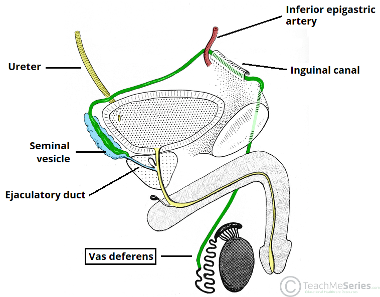
-
Enters the Spermatic Cord
-
Passes through the Inguinal Canal
-
Hooks around the Interfoveolar Ligament and Inferior Epigastric Artery at the Deep Inguinal Ring
-
Crosses the following structures lying on the Obturator Fascia:
- External Iliac Artery and External Iliac Vein
- Obliterated Umbilical Artery
- Obturator Nerve
- Obturator Artery and Obturator Vein
-
Passes across the side wall of the pelvis just under the peritoneum to reach the back of the Bladder
- Throughout its course, no other structure intervenes between it and the peritoneum
-
Crosses the pelvic cavity
-
Curves medially and forwards
-
Crosses above the Ureter
-
Approaches the opposite fellow
-
Two ducts then turn downwards side by side
-
Each dilates in fusiform manner (i.e. becomes the Ampulla)
- The storehouse of Spermatozoa
-
Ampullae lie parallel and medial to the Seminal Vesicles
-
At their lower ends, each loses its thick muscle wall and joins with the outlet of the Seminal Vesicle to form the Ejaculatory Duct
-
Each Ejaculatory Duct passes obliquely though the prostate to open on the Verumontanum
-
Pierces the prostate and opens by the Ejaculatory Duct into the Prostatic Urethra
-
Proximal part of the Vas Deferens absorbs fluid produced by the Seminiferous Tubules of the Testis
- Vas itself only makes a small contribution to the volume of the seminal fluid
-
NB: Lined by Stereo-ciliated Columnar Epithelium