Overview
-
Size: About 8cm x 5cm x 3cm
-
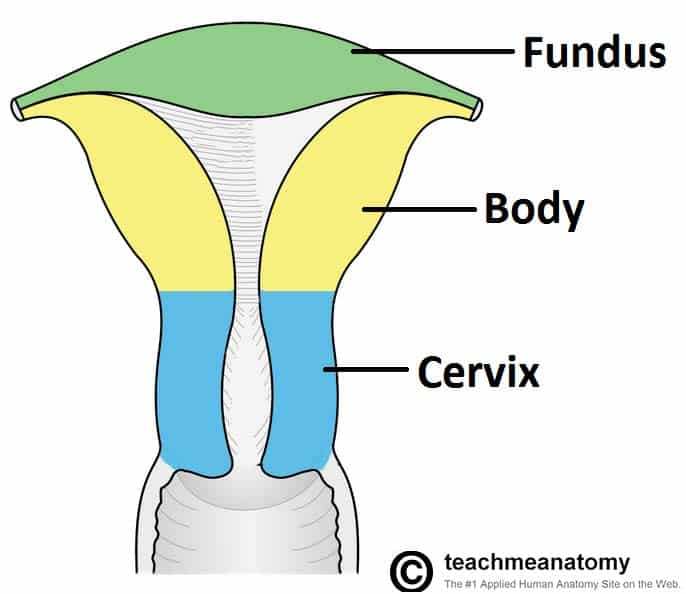
Fundus
- Part above the entrance of the tubes
- Possess a serous coat of pelvic peritoneum which continues over the front and back of the body
-
Body
- Cornu
- Upper angle, at the junction of the Fundus and Body
- Receives the Uterine Tubes
- Enclosed by peritoneum
- Becomes the Broad Ligament laterally
- Intestinal surface – faces upwards with coils of intestine lying upon it
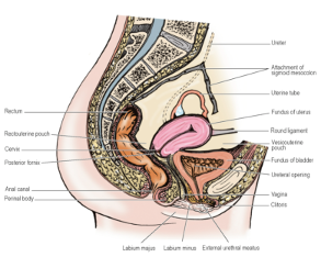
- Vesical surface – faces downwards resting on the Bladder with the peritoneum of the Vesicouterine Pouch intervening
- Cornu
-
Cervix
- Lower end clasped by the vault of the Vagina
- Deep sulcus that surrounds the Cervix = Fornix of the Vagina
- Deepest posteriorly
-
Peritoneum
- Posterior surface covered by peritoneum that continues from the body onto the upper part of the Fornix forming the anterior wall of the Rectouterine pouch (of Douglas)
- NB: Thus, cervix has no direct extra-peritoneal relation to the rectum
- Anterior surface has no peritoneal covering
- Deep to the Vesicouterine Pouch
- Attached to the Bladder above the Trigone by dense connective tissue
- NB: i.e. attached to the Base of the Bladder
- Posterior surface covered by peritoneum that continues from the body onto the upper part of the Fornix forming the anterior wall of the Rectouterine pouch (of Douglas)
-
Ureter is 2cm from the Cervix as it passes first lateral to and then in front of the Fornix
- NB: Uterus body is rarely in the midline – when deviated to one side, the Cervix becomes deflected to the opposite side – thus, one Ureter may be closer to the Cervix
-
Internal Os – the point where the Canal of the Cervix is continuous with the cavity of the body
-
External Os – lower opening into the Vagina
- Circular in the nulliparous
- Transverse slit after childbirth with anterior and posterior lips – anterior lying at a lower level
- On a level with the Ischial Spines
-
NB: Forms an angle of 90 degrees or more with the vagina
Uterine Tubes
- Length: 10cm
- Medial 1cm (intramural part) embedded within Uterine wall
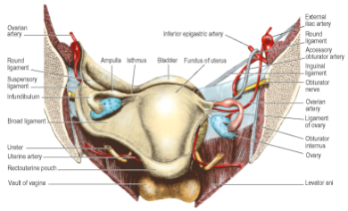
- Lies in the upper edge of the Broad Ligament
- Two layers of smooth muscle – inner circular, outer longitudinal
- Lined by mucous membrane thrown into folds
- Surface epithelium – mixture of ciliated and non-ciliated columnar cells
- Cilia most abundant at fimbriated end (which is least muscular)
- Mesosalpinx – the peritoneal fold embracing the Uterine Tube
- Isthmus:
- The part adjacent to the Uterus
- Straight and narrow
- Ampulla:
- Next to Isthmus
- More than half the length of the tube
- Infundibulum:
- Lateral end of the tube
- Fimbriated end with finger-like processes, the Fimbriae
- Open end lies behind the Broad Ligament adjacent to the lateral pelvic wall
Blood Supply
- Supplies Uterus
- Branch of the Internal Iliac Artery (NB: The anterior division)
- Passes medially across the Pelvic Floor in the base of the Broad Ligament
- Above the Ureter
- Reaches the side of the Supravaginal part of the Cervix
- Gives branch to the Cervix and Vagina
- Turns upwards between layers of the Broad Ligament
- Runs in tortuous manner alongside the Uterus as far as the Cornu
- Gives off branches which penetrate the Uterine walls and anastomose across midline with corresponding branches of the opposite Uterine Artery
- At junction of Uterus and Uterine Tubes, artery turns laterally
- Ends with anastomosing with the Tubal branch of the Ovarian Artery
Ovarian Artery
- Supplies Uterine Tube and Ovary
Veinous Drainage
- Course below the artery at the lower edge of the Broad Ligament
- Form a wide plexus across the pelvic floor
- Communicates with the Vesical and Rectal Plexuses
- Drains to the Internal Iliac Veins
- Tubal Veins join the Ovarian Veins
Lymph Drainage
- From Cervix:
- External and Internal Iliac Nodes
- Sacral Nodes along Uterosacral Ligaments
- Lower part of Uterine Body:
- External Iliac Nodes
- Upper part of Uterine Body, Fundus and Uterine Tube:
- Accompany lymph from Ovaries
- Drain to Para-aortic Nodes
- Few pass to External Iliac Nodes
- Few from region of Uterine Cornua accompany Round Ligaments to reach the Superficial Inguinal Nodes
Nerve Supply
- Branches from the Inferior Hypogastric Plexus
- Smooth muscle of Uterus sensitive to hormonal influences
- Sympathetic supply:
- Vasoconstrictor
- Facilitating function in relation to Uterine muscle
- Division of all Uterine nerves or high transection of the Spinal Cord does not affect Uterine contractility, even in labour
- Pain from Upper Cervix and Body of Uterus (including labour pains)
- Involves T10-L1 – referred to corresponding Dermatomes
- Pre-sacral neurectomy (cutting hypogastric nerves from the Superior Hypogastric Plexus) does not abolish labour pain, but may improve dysmenorrhoea
- Abolition of Uterine sensation requires division of all nerves or transection of the cord above T10
- Pain from Cervix:
- Carried by Pelvic Splanchnic Nerves
- Pain from Upper Cervix appears to run with Sympathetic nerves
- Cervix and Body relatively insensitive to cutting and burning
- Uterine Tube sensitive to touching and cutting
Structure
Myometrium
- Smooth muscle
- Bulk of the Uterus
- Fibres in 3 layers but ill-defined
- Outer longitudinal and expulsive
- More deeply placed are circular and act as sphincters round the larger blood vessels, the openings of the Uterine Tubes and the Internal Os
Endometrium
- Columnar epithelium that dips down into the endometrial stroma to form the endometrial glands
- At menstruation, the bases of the glands remain to provide the source for the new epithelial covering
- Mucosa of Cervix does not take part in the cyclical changes and is not shed at menstruation
- Surface cells are mucus-secreting and there are also mucous glands
- Just inside the External Os the epithelium changes to the Stratified Squamous epithelium of the Vagina
- Outer or serous covering of the Uterus is the Peritoneum
Supports
- Normal position of Uterus: Anteflexion and Anteversion
- External os thus opens through the anterior wall of the Vagina
- 20% of nulliparous females may have a retroverted Uterus
- Most fixed part of Uterus is Cervix
- Pubovaginalis part of Levator Ani and the Perineal Body support the Vagina
- Assists indirectly in holding the Cervix up
- If muscles unduly stretched or damaged during childbirth, posterior Vaginal wall sinks downwards (prolapses) and this often followed by prolapse or retroversion of the Uterus
- NB: Thus, it can be said the uterus is supported by the Levator Ani muscle
Broad Ligament
- Lax double fold of Peritoneum
- Lies lateral to the Uterus
- Plays little part in uterine support
- Medial edge attached to side wall of Uterus
- Flows over its intestinal and vesical surfaces as its serous coat
- Lateral edge attached to side wall of pelvis
- Line of lateral attachment crosses:
- Obturator Nerve
- Superior Vesical or Obliterated Umbilical Vessels
- Obturator Artery and Vein
- Line of lateral attachment crosses:
- Upper border
- Free
- Forms Mesosalpinx
- Contains Uterine Tube
- Upper lateral part contains Ovarian Vessels and Lymphatics
- Extended over the External Iliac Vessels as a fold – Suspensory Ligament of the Ovary
- Two layers of inferior edge pass forwards and backwards to line pelvic cavity
- Ureter adhering underneath
- Anterior layer of Broad Ligament bulged forwards by Round Ligament of Uterus just below the Uterine Tube
- Posterior layer has a fold projecting backwards suspending the ovary – Mesovarium
- Between layers of Broad Ligament is a mass of areolar tissue – Parametrium
- Lies the Uterine Vessels
- Lymphatics
- Round Ligament of Uterus
- Ligament of the Ovary
- Vestigial remnants of Mesonephric Tubules
Round Ligament of Uterus
- Comprised of smooth muscle and fibrous tissue
- Holds the Uterus forwards in anteflexion and anteversion – esp. when forces tend to push the Uterus backwards, e.g. bladder distension, gravity during recumbency
Course:
- Extends from junction of Uterus and Tube to Deep Inguinal Ring
- Lies in Broad Ligament below Uterine Tube
- Bulges anterior layer of the Broad Ligament forwards
- Round Ligament passes through Inguinal Canal
- Attached at distal extremity to fibrofatty tissue of the Labium Majus of the Vulva
- Continuous with Ligament of the Ovary (through its uterine attachment)
- Two ligaments together represent the Gubernaculum
- Arterial Supply:
- Branch of Ovarian Artery in the Broad Ligament
- Branch of Inferior Epigastric Artery in the Inguinal Canal
Transverse Cervical Ligament
- A.k.a. Lateral Cervical, Cardinal or Mackenrodt’s Ligament
- Thickenings of connective tissue in the base of each Broad Ligament
- Extends from Cervix and Vaginal Fornix laterally to the side wall of the Pelvis
- Following traverse the connective tissue of this ligament:
- Ureter
- Uterine Artery
- Inferior Hypogastric Plexus
- Imparts lateral stability to the Cervix and an important support of the Uterus
- Uterosacral Ligaments:
- Fibrous tissue and smooth muscle
- Extend backwards from the Cervix below the peritoneum
- Embraces the Rectouterine Pouch and Rectum
- Becomes attached to the front of the Sacrum
- Palpable on rectal exam
- Keep Cervix braced backwards against forward pull of the Round Ligaments on the Fundus
- Maintains the body of Uterus in anteversion
Development
- Paramesonephric (Mullerian) Ducts develop as a linear invagination of the Coelomic Epithelium on the lateral aspect of the Mesonephros
- Grow caudally lateral to the Mesonephric Ducts
- Cross ventral to them and fuse at their caudal ends to make the Uterus
- Continue to reach the dorsal wall of the Urogenital Sinus – forms the upper part of the Vagina
- Cranial ends persist as the Uterine Tubes
- Incomplete fusion results in a Median Septum in the Uterus or a Bicornuate Uterus
Surgical Approach
- Total Hysterectomy:
- Broad, Round and Ovarian Ligaments and Uterine Tubes divided on each side near the Uterus
- Lower ends of Ureters need to be safeguarded esp. when Uterine Arteries are divided
- Anterior and Posterior Vaginal walls cut across below the Cervix
- If Subtotal Hysterectomy via abdominal route:
- Cervix is cut across the level of the Lateral Ligaments without opening into the Vagina