Section: Colorectal
Definition
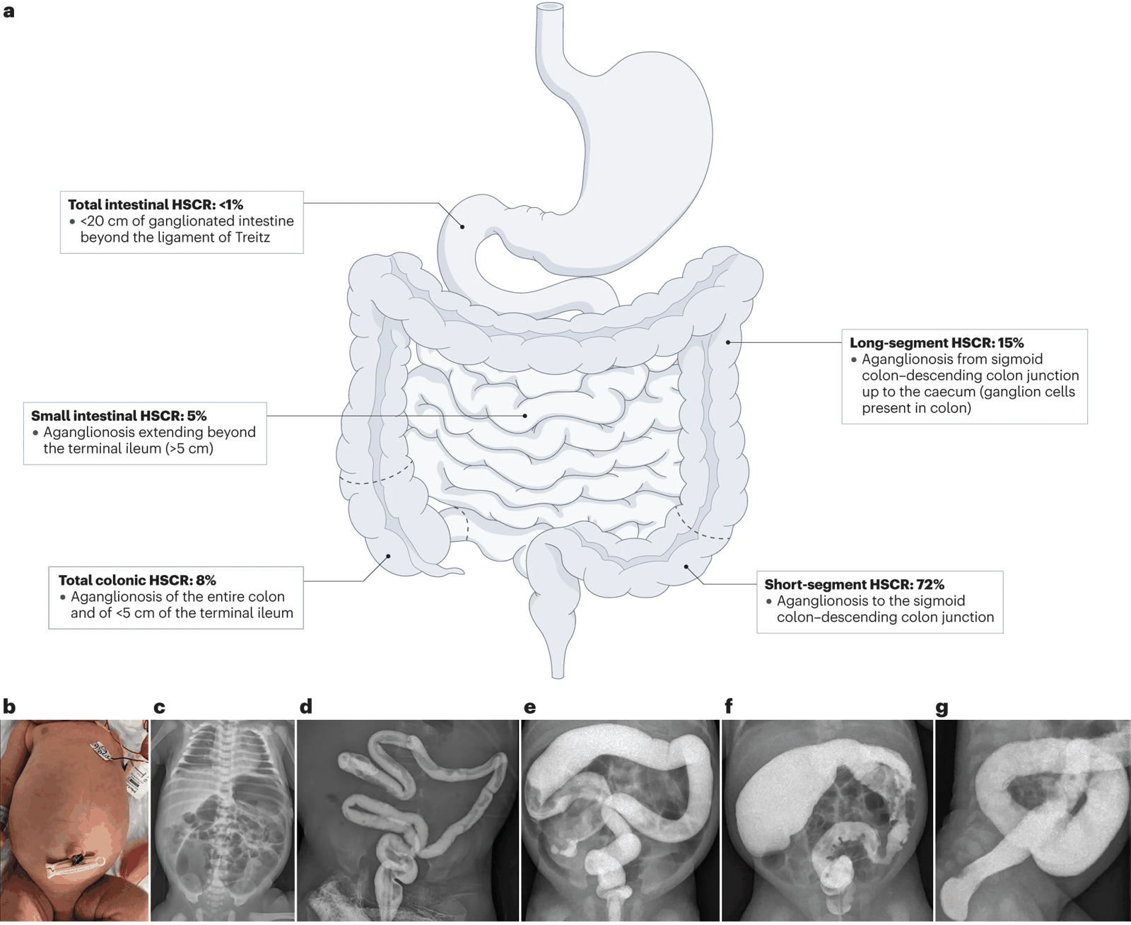
- Congenital disorder characterised by absence of ganglion cells in the distal rectum extend a variable distance proximally.
- Affects:
- The myenteric plexus between the longitudinal and circular muscle layers (Auerbach plexus)
- The submucosal plexus between the mucosal and circular muscle layers (Meissner plexus)
- Affects:
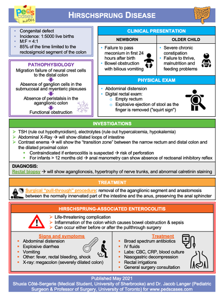
- Epidemiology: 1/5,000, M : F = 4 : 1
Aetiology
- Disturbed migration of neural crest cells along variable length of intestine.
- Under control of RET gene.
- Causes total absence of ganglion cells in the nerve plexuses.
- Causes overactivity of intestine with persistent release of acetylcholine, with continuous contraction of the narrowed (affected) segment of colon, with progressive secondary dilatation of healthy proximal colon.
Clinical
- History: constipation or overflow, abdominal swelling, feeding difficulties
- Examination: rectal exam – may have absence of normal relaxation of anal sphincter muscles
Investigations
- Imaging: may be useful to assess for structure/function and identify obstruction of colon
- Biopsy: rectal biopsy taken which checks for presence/absence of ganglion cells with hypertrophy of nerve fibres in the aganglionic segment
- Anorectal manometry may be useful
Surgery
May need temporary decompressive colostomy prior to definitive pull-through procedure
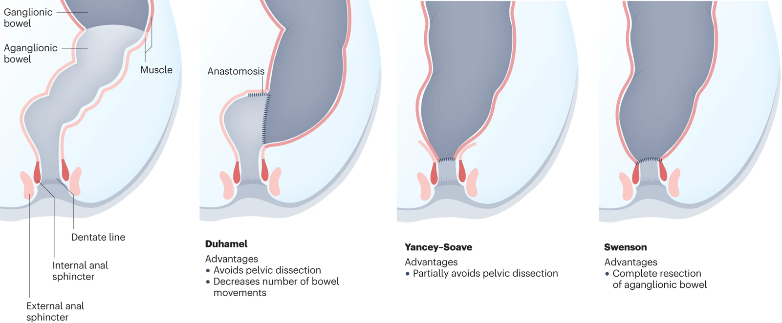
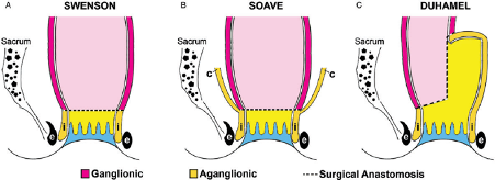
Swenson procedure
- a radial full-thickness resection is begun in the aganglionic rectum just proximal to the dentate line, and the aganglionic segment and transition zone are pulled through the anus and resected. The transected end of ganglionic bowel proximal to the aganglionic segment is sutured end to end to the terminal rectum. Soave procedure
- the initial radial incision is made through the mucosa, and the dissection is extended proximally in the submucosal plane for several centimetres before dividing the muscularis propria. As a consequence, the muscularis propria of the distal rectum is retained as a ‘‘cuff’’ (c), through which the ganglionic bowel is drawn inferiorly and attached just above the dentate line. Duhamel procedure
- the pull-through is performed along the posterior aspect of the aganglionic rectum, and ganglionic bowel is attached in a side-to-side manner to the posterior rectum. The anterior rectum remains as an aganglionic pouch.