Section: Colorectal Sub-section: Proctology Curriculum: Curriculum, page 19
Definition
- Involuntary loss of solid, liquid or gas, with varying debilitating effect on lifestyle.
- Passive
- Lack of awareness
- Urge
- incontinance despite effort to retain stool
The ability to maintain continence depends on: - Stool consistency
- Rectal capacity and compliance
- Neural pathways
- Anal sphincter and pelvic floor function
- Anorectal sensation
Anatomy
Mechanical or anatomical factors
Anal sphincter
- Composed of internal and external sphincter.
- Internal anal sphincter – expansion of the circular layer of the bowel wall. Tone contributes 70-80% of the resting sphincter pressure.
- External anal sphincter – extension of the levator ani muscle. Contributes to the ability to voluntarily squeeze the anal sphincter complex.
- Thus, passive leakage is usually a problem with the internal sphincter, whera’s frank incontinence and urge symptoms are due to the external sphincter.
Anal mucosal folds and vascular cushions
- Help to create a seal and contribute about 10% of the resting tone
Puborectalis muscle
- Forms a sling around the rectum which leads to the anal-rectal angle which acts like a flap valve.
Rectal and anal sensation
External anal sphincter
- Is innervated by the Pudendal Nerve (S2-S4)
Puborectalis muscle
- is innervated by pelvic branches of S3-S4
Parasympathetic nerves
- from S2-S4 via the nervi erigentes.
- Rectal distention is transmitted by this pathway.
Sympathetic nerves
- Come from the 5th Lumbar nerve root via the superior and inferior hypogastric plexus.
All of the above contribute the maintenance of continence. The anorectal inhibitory reflex pathway is when the rectum is full, the internal anal sphincter relaxes, which leads to a reflexive contraction of the external anal sphincter. This provides a sensation of urgency, and leads to relaxation of the puborectalis muscle which decreases the angulation of the rectum and allows defectation.
Rectal compliance
- Rectum is a reservoir which allows stool storage.
- If compliance is altered – can cause incontinence.
Stool consistency
- Self explanatory.
Aetiology
3 main mechanisms:
- Diarrhoea/loose stool
- Diseases causing diarrhoea
- Overflow from constipation
- Rectal volume/compliance
- Sphincter saving procedures
- IBD
- Radiation proctitis
- Anatomical or functional injury to anal sphincter complex
- Previous anal operations:
- Internal sphincterotomy
- Fistula surgery
- Haemorrhoidectomy
- Manual dilatation of the anus
- Childbirth
- Previous hysterectomy
- Procidentia
- Rectal prolapse
- Trauma
- Carcinoma
- Irradiation
- Neurogenic causes – CNS, spinal, diabetic Can be idiopathic or multifactorial.
- Previous anal operations:
Clinical
History:
- Stool diary for frequency and severity of incontinence episodes.
- Stool consistency using Bristol stool chart.
- QoL assessments e.g., Faecal Incontinence Quality of Life instrument.
- Associated urinary incontinence, constipation
- Urge (may be related to external sphincter) vs passive/insensible
- PMHx: Obstetric history, previous anal surgery, diabetes, radiation Examination:
- General – abdomen, neurological exam of back and lower limbs
- Perianal skin inspected for scars (trauma or surgery), excoriation.
- Open anal orifice suggests decreased resting tone, weak voluntary contraction and pudendal neuropathy
- Asked to strain to accentuate descending perineum or exteriorise rectal prolapse
- Sensory perception at anal margin
- DRE to assess: resting and squeeze pressures, contraction of puborectalis, check for rectocele, defects in sphincter, impacted stool.
Investigations
Depends on duration, severity, response to initial conservative management and fitness for surgery.
Exclude organic pathology
- Colonoscopy (or flexi sig)
- Pelvic ultrasound, cervical smears
Anorectal physiology
- Manometry
- Measures internal (resting) and external sphincter (squeeze) tone
- Anal and rectal sensation testing
- Most common is electrical sensitivity testing
- Pudendal nerve terminal latency (PNTL) - not used anymore
- Measured with a finger-mounted St. Mark’s electrode, which stimulates the pudendal nerve at the level of the ischial spine and records the conduction time to the sphincter. Normal PNTL is less than 2.2 milliseconds and prolongation is intended to reflect nerve damage.
Imaging
- Endoanal USS
- Dynamic assessment of thickness and structure of external and internal sphincter muscle
- First line to diagnose sphincter defects w suspected injuries
- Standard or MRI dynamic defecography
- Useful in select cases where mixed symptoms including obstructive defecation, to identify an occult prolapse
Management
MDT approach. Guided by severity, aetiology and structural integrity of sphincter muscles.
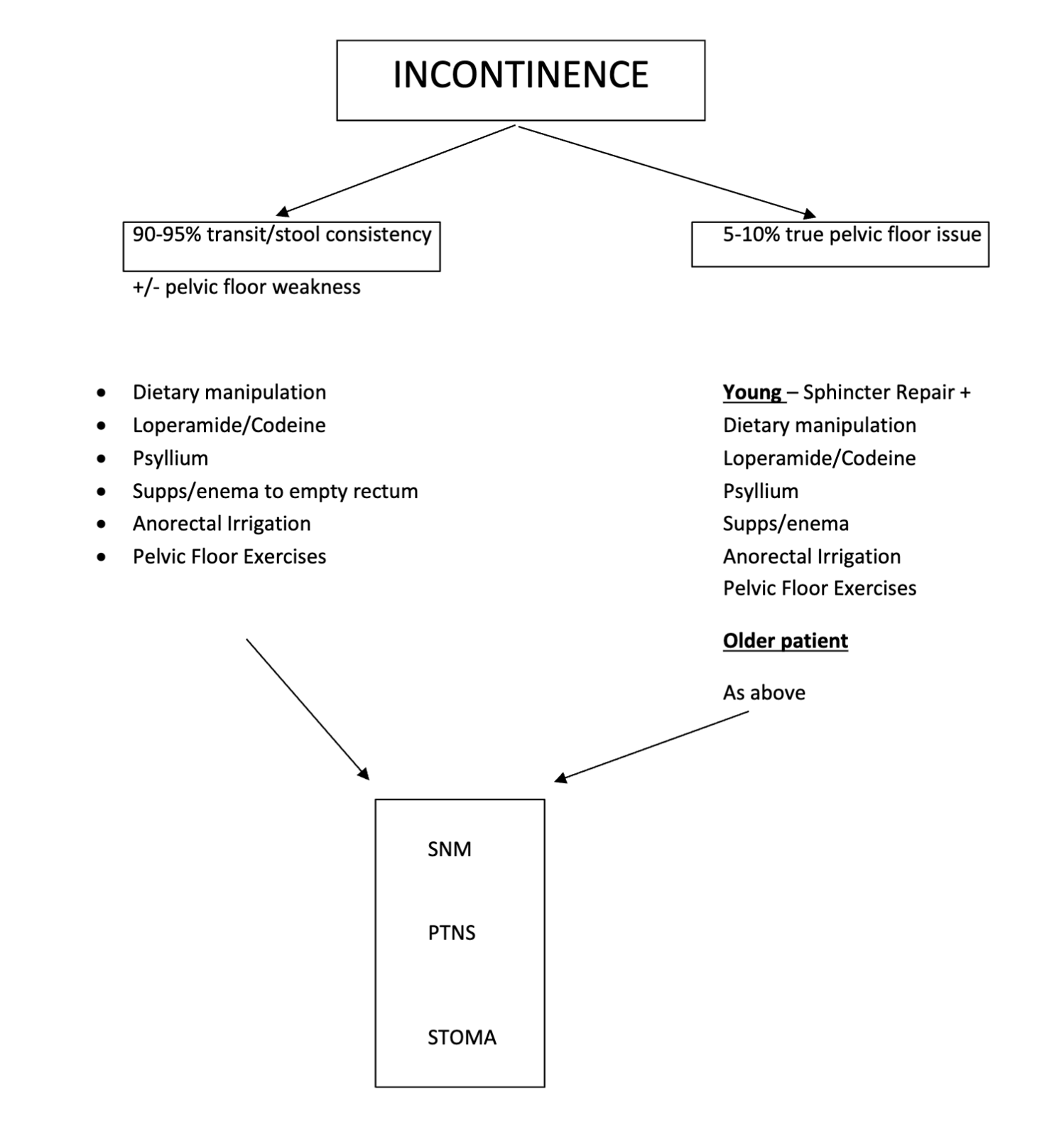
Remember 90-95% of cases of FI are due to a colonic problem – transit/stool consistency and only 5% -10% are due to true sphincter/pelvic pathology
Conservative
- Dietary modification and medications
- Increase in fibre may improve consistency
- PSYLLIUM – can be used as a bulking agent either alone or in combination with Loperamide
- Enemas/suppositories to allow complete evacuation
- Antidiarrhoeal agents – loperamide, codeine, cholestyramine if bile salt related
- Loperamide MOA: opioid. Mu-receptor agonist. Acts directly on circular and longitudinal intestinal muscles to reduce transit, inhibit peristalsis and increase rectal tone.
- Take 30 minutes before meals EVERY DAY. This becomes the regular daily dose (explain clearly to pts the need to take daily and that is very safe long term)
- Biofeedback and pelvic floor muscle retraining
- Behavioural therapy
- When combined with electrical stimulation, may be more effective than either alone.
- Anal plug
- Disposable device that expands when soaked with rectal mucus and controls continence by blocking passage of stool. Poorly tolerated.
- ANORECAL IRRIGATION
- SACRAL NEUROMODULATION (SNM)
- POSTERIOR TIBIAL NERVE STIMULATION (PTNS)
Surgical
Direct repair of sphincter
- Sphincteroplasty
- Anterior anal sphincter repair:
- Transverse incision between anus and vaginal introitus.
- Scar tissue and muscle ends dissected with adequate mobilisation necessary to ensure tension free.
- Scar tissue is divided, two muscle ends overlapped over midline with 2/0 mattress.
- T-closure of skin with absorbable sutures
- Anterior anal sphincter repair:
- Techniques to augment sphincter function
- Injectables
- Tibial nerve stimulation
- Sacral nerve and
- Alteration of ascending spinal sensory pathways
- Minimally invasive, minimal morbidity, expensive. Variable outcome.
- Diagnostic phase of peripheral nerve evaluation, under LA or GA. S3 foramen cannulated under fluoroscopy with electrode with stimulation performed looking for appropriate ‘bellows response’ of pelvic floor and plantar flexion of ipsilateral great toe. Then 3-week trial of stimulation while filling out bowel habit diary.
- Therapeutic phase, if reduction by > 50% on diagnostic phase. Permanent stimulator placed subcutaneously in gluteal area under LA.
- Contraindications: sacral pathology, severe sphincter damage, pregnancy, bleeding risk, psychological instability, pacemaker or ICD
- Replace sphincter complex
- Muscle transposition (rare)
- Gluteal muscle or gracilis, wrapped around anus to form new sphincter.
- Artificial sphincter (rare)
- Muscle transposition (rare)
- Stoma or antegrade continence enema (e.g. appendicostomy or CHAIT)