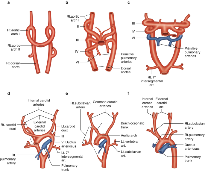
The aortic arch embryology involves the transformation of six paired pharyngeal (branchial) arch arteries that develop sequentially and remodel into the major arteries of the head, neck, and thorax. These arches form from the mesoderm around the developing pharyngeal apparatus.
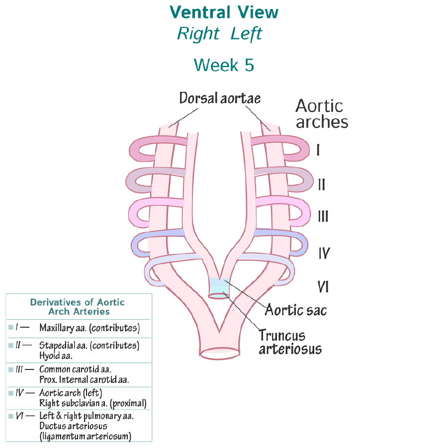
- 1st arch
- Mostly regresses
- Remnant forms part of maxillary artery
- 2nd arch
- Mostly regresses
- Remnant contributes to the stapedial and hyoid arteries
- 3rd arch
- Forms the common carotid arteries
- Also forms the proximal internal carotid arteries
- 4th arch
- Left side: forms part of the aortic arch (between left common carotid and left subclavian)
- Right side: forms the proximal right subclavian artery
- 5th arch
- Either does not form or forms transiently and regresses completely (has no adult derivative)
- 6th arch (pulmonary arch)
- Proximal parts form the right and left pulmonary arteries
- Distal part of left 6th arch becomes the ductus arteriosus, which later becomes the ligamentum arteriosum
- Distal right 6th arch regresses
- Dorsal aortae
- Initially paired, they fuse to form the descending thoracic and abdominal aorta
- Segments contribute to vertebral, intercostal, and other arteries
- Double aortic arch
- Failure of regression of the right dorsal aorta
- Forms a vascular ring that encircles and compresses the trachea and esophagus
- Symptoms: stridor, dysphagia, respiratory distress in infancy
- Right aortic arch
- Regression of the left dorsal aorta with persistence of the right
- Aortic arch runs on the right side of the trachea and esophagus
- Can be isolated or associated with vascular rings or congenital heart disease (e.g., Tetralogy of Fallot)
- Aberrant right subclavian artery (Arteria Lusoria)
- Right 4th arch and proximal right dorsal aorta regress
- Right subclavian arises from the distal aortic arch (after left subclavian), passes posterior to the esophagus
- Usually asymptomatic but may cause dysphagia lusoria if compressive
- Aberrant left subclavian artery (in setting of right-sided aortic arch)
- Analogous to ARSA, left subclavian arises abnormally and may course behind the trachea/esophagus
- Interrupted aortic arch
- Segment of aortic arch (commonly the left 4th arch) fails to develop
- Classified into types A, B, and C depending on location of interruption
- Associated with DiGeorge syndrome (22q11.2 deletion) and other congenital heart defects
- Coarctation of the aorta
- Narrowing of the aortic lumen, commonly near the ductus arteriosus (juxtaductal)
- Can be pre-ductal or post-ductal
- Often associated with bicuspid aortic valve and Turner syndrome
- Persistent ductus arteriosus (PDA)
- Failure of closure of the left distal 6th arch derivative after birth
- Allows continued shunt from aorta to pulmonary artery, leading to left heart volume overload if significant