- Circulatory Pathways
- Oxygenated Blood from Placenta:
- Oxygen-rich blood flows from the placenta to the fetus via the umbilical vein.
- The blood passes through the liver via the ductus venosus to the inferior vena cava (bypassing most of the liver).
- Ductus Venosus:
- Allows blood to bypass the liver, entering the inferior vena cava and mixing with deoxygenated blood.
- Inferior Vena Cava:
- Carries mixed blood into the right atrium of the heart.
- Right Atrium to Left Atrium (Foramen Ovale):
- Blood moves from the right atrium to the left atrium through the foramen ovale, bypassing the lungs.
- Left Atrium to Left Ventricle:
- Oxygenated blood flows from the left atrium to the left ventricle, then into the aorta for systemic circulation.
- Right Ventricle and Pulmonary Artery:
- Blood from the right atrium moves into the right ventricle and then is pumped into the pulmonary artery.
- Blood is diverted through the ductus arteriosus to the aorta, bypassing the lungs.
- Ductus Arteriosus:
- Connects the pulmonary artery to the aorta, allowing blood to bypass the lungs.
- Oxygenated Blood to Head and Upper Body:
- Oxygenated blood from the aorta is distributed to the head, neck, and upper limbs.
- Deoxygenated Blood:
- Deoxygenated blood from the body returns to the fetus through the superior vena cava, mixing in the right atrium to continue the cycle.
- Shunts in Fetal Circulation
- Foramen Ovale:
- Allows blood flow from the right atrium to the left atrium, bypassing the lungs.
- Ductus Arteriosus:
- Connects the pulmonary artery to the aorta, bypassing the lungs.
- Ductus Venosus:
- Diverts blood from the umbilical vein directly to the inferior vena cava, bypassing the liver.
- Postnatal Changes After Birth
- Lung Function:
- After birth, the lungs expand and blood flow to the pulmonary circulation increases.
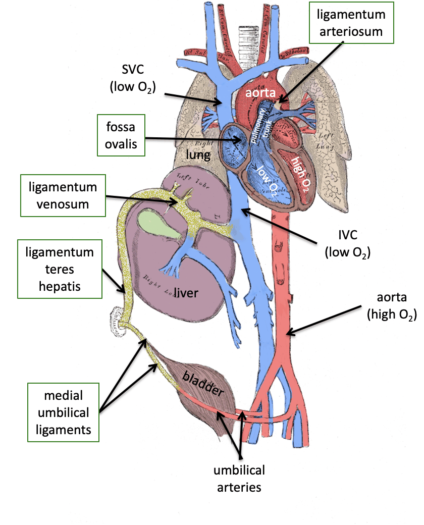
- The ductus arteriosus, ductus venosus, and foramen ovale close after birth.
- Closure of Shunts:
- Ductus arteriosus: Closes within 24-48 hours, becoming the ligamentum arteriosum.
- Foramen ovale: Closes functionally shortly after birth, becoming the fossa ovalis.
- Ductus venosus: Closes and becomes the ligamentum venosum.
- Clinical Relevance
- Patent Ductus Arteriosus (PDA):
- Failure of the ductus arteriosus to close after birth, causing abnormal blood flow between the pulmonary artery and aorta.
- Patent Foramen Ovale (PFO):
- A persistent opening between the right and left atria that can lead to stroke or paradoxical embolism.
- Congenital Heart Defects
- Abnormalities in fetal circulation pathways can lead to conditions such as cyanotic congenital heart defects or truncus arteriosus.