Gonadal Genesis
See: Mal-descent of the testis
Overview
- Three structures comprise the gonad:
- Primordial Germ Cells - Migrate to Gonadal Ridge from Yolk Sac
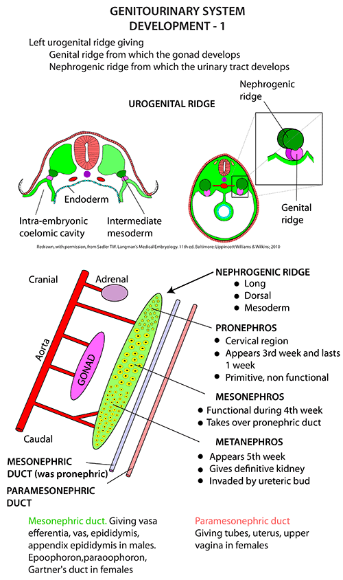
- Genital Ridge (Mesenchyme of mesonephros)
- Coelomic Epithelium
- Steps overview
- 4 weeks - formation of genital ridge
- 6 weeks - migration of primordial germ cells
- 7 weeks - Differentiation into testes of ovaries (SRY gene)
- 8-10 weeks - Gonadal development
- 10-12 weeks - Maturation
Weeks 4-6
- Initial Development and Formation of Genital Ridges
- Genital Ridge Formation
- Around week 4, the genital ridges (future gonads) begin to form along the mesonephros in the intermediate mesoderm.
- These undifferentiated ridges will later give rise to either testes or ovaries.
- Around week 4, the genital ridges (future gonads) begin to form along the mesonephros in the intermediate mesoderm.
- Primordial Germ Cells Migration
- Primordial germ cells (PGCs), which are precursors to gametes, migrate from the yolk sac to the genital ridges by week 6, settling in the developing gonads.
- Genital Ridge Formation
Week 7
- Differentiation into Testes or Ovaries
- Genetic Determination
- The genetic sex (XX for female, XY for male) begins to influence the differentiation pathway.
- SRY Gene Activation (if XY)
- In the presence of the SRY (Sex-determining Region Y) gene, the undifferentiated gonads start developing into testes.
- Sertoli cells begin to form and produce Anti-Müllerian Hormone (AMH), which suppresses the development of female reproductive structures.
- Absence of SRY (if XX)
- In the absence of the SRY gene, the gonads develop into ovaries.
- There is no AMH production, allowing the Müllerian ducts to persist, which will form female reproductive structures (fallopian tubes, uterus, upper vagina).
- Genetic Determination
Weeks 8-10
- Testis Development (if XY):
- Sertoli cells further differentiate, organizing around the germ cells to form testis cords, which will later develop into seminiferous tubules.
- Leydig cells appear and start producing testosterone, promoting the development of male external genitalia and the Wolffian duct system (epididymis, vas deferens, and seminal vesicles).
- Ovary Development (if XX):
- The absence of AMH allows the Müllerian ducts to continue developing.
- Oogonia (female germ cells) start proliferating and differentiate into primary oocytes within developing ovarian follicles.
Weeks 10-12
- Further Maturation
- Male Gonads (Testes)
- By week 10, testosterone production helps masculinize external genitalia, while AMH ensures regression of the Müllerian ducts.
- Female Gonads (Ovaries)
- By week 12, oogonia cease proliferation and enter meiosis, halting at the primary oocyte stage, which will remain dormant until puberty.
- Male Gonads (Testes)
Supporting structures
Mesonephric duct (Wolffian duct)
- Derivates of Mesonephric Duct
- Canal of Epididymis
- Vas Deferens
- Ejaculatory Duct
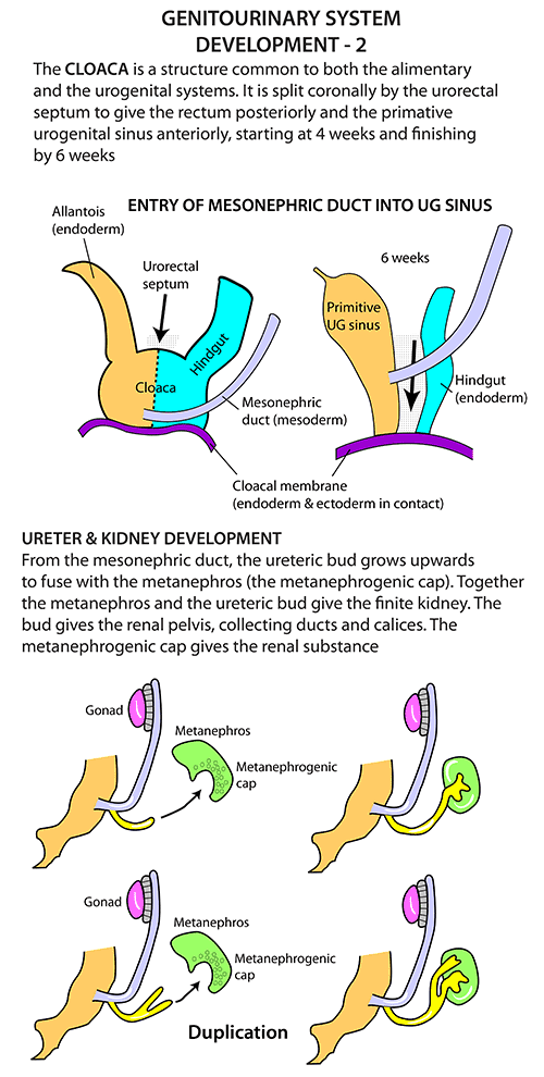
- The mesonephric ducts develop alongside the mesonephros (an early kidney structure)
- In males, the mesonephric ducts are preserved and differentiate into reproductive structures under the influence of testosterone produced by Leydig cells and Anti-Müllerian Hormone (AMH) produced by Sertoli cells.
Paramesonephric duct (Müllerian duct)
- Forms
- Fallopian Tubes: The upper portions of the ducts form the fallopian tubes.
- Uterus: The middle portions of the ducts fuse at the midline to form the uterus.
- Cervix and Upper Vagina: The lower portions of the fused ducts become the cervix and the upper part of the vagina.
- In men
- Appendix Testis
Mesonephric Tubules
- Derivates of remaining Mesonephric Tubules
- Vasa Efferentia of Testis
- Paradidymis
Mesorchium
- Double peritoneal fold suspending the testis
- Upper fold transmits spermatic vessels
- Lower fold: Gubernaculum
- Gubernaculum
- Condensation of Mesodermal cells connecting lower pole of Testis to region of anterior abdo wall that layer forms the Scrotum
- Abnormal insertion of the Gubernaculum
- Leads to ectopic testis (cf undescended testis or cryptorchidism
- Gubernaculum
Processus Vaginalis
- Sac of peritoneum that protrudes down the inguinal canal anterosuperior to the Gubernaculum
Gonadal Descent
Phase 1: Transabdominal Descent (Weeks 8-15)
- Descent Toward the Inguinal Region
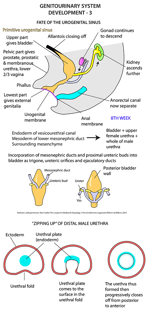
- Week 8 - testes are in the upper abdomen near the kidney.
- Weeks 10 and 15 - begin descending toward the inguinal region
- Pulled down by the shortening of the gubernaculum and influenced by anti-Müllerian hormone (AMH) from Sertoli cells
- Gubernaculum – a condensation of mesodermal cells connecting the lower pole of the Testis to the region of the anterior abdominal wall that later forms the Scrotum
- Traverses the site of the future Inguinal Canal
- Pulled down by the shortening of the gubernaculum and influenced by anti-Müllerian hormone (AMH) from Sertoli cells
- Peritoneum and Processus Vaginalis
- The peritoneum forms a pouch, the processus vaginalis, which follows the gubernaculum into the inguinal canal. This will eventually become part of the covering around the testes in the scrotum.
Phase 2: Inguinoscrotal Descent (Weeks 25-35)
- Descent through the Inguinal Canal:
- Week 25 - testes start their journey through the inguinal canal.
- This phase is androgen-dependent, relying on testosterone production from Leydig cells.
- Week 25 - testes start their journey through the inguinal canal.
- Final Descent into the Scrotum:
- Weeks 33-35 - the testes have usually descended fully into the scrotum.
- The processus vaginalis typically closes, leaving a small serous pouch (the tunica vaginalis) around each testis in the scrotum.
- 50-75% of infants have complete closure at birth
- Most of the remaining will close over the next 12 months
- Failure of obliteration of processus vaginalis leads to
- Hydrocele, Hernia, Encysted hydrocele of the cord
- Right testis descends later than left
- ↑ Incidence of abnormalities on R side
- ↑ Incidence of hernias in premature babies
- Normal post-partum intra-abdominal pressures make it more difficult for the processus to close spontaneously
- The processus vaginalis typically closes, leaving a small serous pouch (the tunica vaginalis) around each testis in the scrotum.
- Weeks 33-35 - the testes have usually descended fully into the scrotum.