- Derived from Mesoderm
- In early embryogenesis, the mesoderm forms three parts
- Paraxial mesoderm → forms somites (gives rise to axial skeleton, skeletal muscles, dermis)
- Intermediate mesoderm → forms the urogenital structures
- Lateral plate mesoderm → forms body wall, limbs, circulatory system
- The intermediate mesoderm specifically develops into:
- Nephrogenic cord → forms kidneys (pronephros, mesonephros, metanephros)
- Gonadal ridge → forms ovaries or testes
- Also gives rise to ureters, parts of genital ducts, and adrenal cortex (partly)
- Intermediate mesoderm
- Gives rise to the urogenital ridge, which forms along the dorsal body wall
- Divides into:
- Nephrogenic cord → urinary system
- Gonadal ridge → reproductive system
- These two regions develop in close proximity and influence each other structurally and hormonally
- Kidney development
- Occurs in three successive stages, all derived from the nephrogenic cord:
- Pronephros (week 4): rudimentary, non-functional; regresses quickly
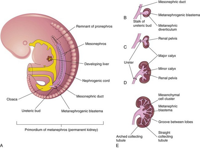
- Mesonephros (late 4th to 6th week):
- Functions temporarily as an embryonic kidney
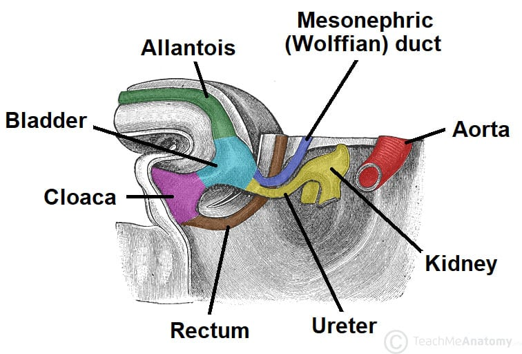
- Produces urine that drains via mesonephric (Wolffian) ducts into the cloaca
- Also provides ductal infrastructure that will later support gonadal development
- Metanephros (starts week 5; functional by week 10):
- Forms the definitive kidney
- Arises from two interacting components:
- Ureteric bud: sprouting from the mesonephric duct → forms ureter, renal pelvis, calyces, collecting ducts
- Metanephric mesenchyme: interacts with bud → induces nephron formation (glomeruli to distal tubule)
- Migration: kidneys ascend from the pelvis to the lumbar region, rotating medially
- Formation of ureters and bladder
- As kidneys ascend, the ureteric buds separate from the mesonephric ducts and attach directly to the developing bladder
- Bladder and urethra develop from the urogenital sinus, derived from the division of the cloaca by the urorectal septum
- The trigone of the bladder develops from incorporated distal mesonephric ducts
- The allantois (from urogenital sinus) becomes the urachus, which should obliterate to become the median umbilical ligament
- Gonadal development and relationship to urinary tract
- Gonads arise adjacent to the mesonephros, and early support structures are derived from the same embryonic tissues
- Primordial germ cells migrate from the yolk sac into the gonadal ridge
- Indifferent gonads can develop into testes or ovaries based on genetic and hormonal signals
- SRY gene (Y chromosome) → testes → produce:
- AMH (anti-Müllerian hormone) → regression of Müllerian (paramesonephric) ducts
- Testosterone → stimulates Wolffian (mesonephric) ducts → epididymis, vas deferens, seminal vesicles
- No SRY (XX genotype) → ovaries → lack AMH/testosterone →
- Persistence of Müllerian ducts → fallopian tubes, uterus, upper vagina
- Regression of mesonephric ducts
- Integration and descent
- Gonads remain connected to early kidney structures (mesonephros) via mesonephric tubules and ducts
- In males, the mesonephric ducts are retained and become part of the reproductive tract
- Gonads (especially testes) descend along a path influenced by the gubernaculum and associated peritoneum, passing through the inguinal canal
- So, the nephric and gonadal systems share:
- A common embryologic origin (intermediate mesoderm)
- Close spatial development, especially early on
- Intertwined ductal systems, where the mesonephric duct plays a dual role in early kidney function and later in male reproductive tract development
- Renal agenesis
- Cause: Failure of the ureteric bud to form or interact properly with the metanephric mesenchyme (metanephric blastema).
- Embryological link: Both structures arise from intermediate mesoderm; if the ureteric bud doesn’t grow from the mesonephric duct or fails to induce the metanephric mesenchyme, the kidney won’t form.
- Unilateral: Usually asymptomatic with compensatory hypertrophy.
- Bilateral: Incompatible with life (leads to Potter sequence).
- Renal hypoplasia
- Cause: Incomplete development of nephrons due to reduced interaction between ureteric bud and metanephric mesenchyme.
- Link: Suggests impaired signaling within the intermediate mesoderm derivatives.
- Ectopic kidney / Horseshoe kidney
- Cause: Abnormal migration of the kidney during development. Kidneys form in the pelvis and ascend.
- Horseshoe: Inferior poles fuse and get trapped under the inferior mesenteric artery.
- Link: Reflects miscoordination in positional development of intermediate mesoderm structures.
- Multicystic dysplastic kidney (MCDK)
- Cause: Abnormal induction of metanephric mesenchyme by the ureteric bud — leading to cystic, non-functional tissue.
- Link: Again, failed reciprocal signaling between ureteric bud and metanephric blastema.
- Duplex kidney / ureters
- Cause: Two ureteric buds form or early bifurcation of the ureteric bud.
- Link: Overactivity of mesonephric duct–derived tissue (ureteric bud) in intermediate mesoderm.
- Posterior urethral valves (PUV)
- Cause: Obstruction in the posterior urethra due to persistent membranes.
- Embryo link: Abnormal development of the cloaca and urogenital sinus (partially from endoderm, but linked closely with intermediate mesoderm derivatives).
- Effect: Causes bladder outflow obstruction → bilateral hydronephrosis → renal dysplasia.
- Urachal anomalies (e.g., patent urachus, urachal sinus/cyst)
- Cause: Failure of involution of the allantois–urachus tract connecting the bladder to the umbilicus.
- Embryo link: Though not directly from intermediate mesoderm, closely associated with urogenital sinus (which develops in parallel).
- Polycystic kidney disease (PKD)
- Cause: Genetic defect affecting tubular development → cyst formation.
- Embryo link: Affects the epithelial cells lining collecting ducts, derived from the ureteric bud (intermediate mesoderm origin).