- Longitudinal channels develop to return blood to the Sinus Venosus
- Receives blood from 3 sources:
- Placenta via Umbilical Veins
- Yolk Sac via Vitelline Veins
- General Tissue of Embryo via Cardinal Veins
- Each group has Right and Left veins with anastomosing cross-channels between each pair
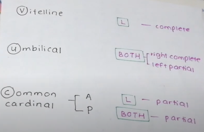
- All or part of one longitudinal vein from each pair disappears:
- Right Umbilical Vein
- Left Vitelline Vein
- Left Cardinal Vein
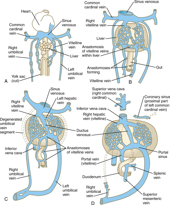
Regression
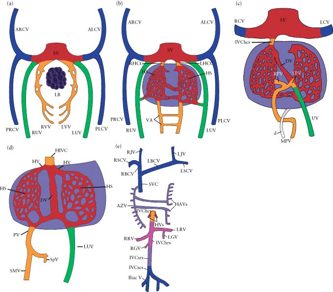
Vitelline Veins
- The right and left vitelline veins run from the yolk sac, passing through the septum transversum, and drain into the sinus venosus of the primitive heart.
- They form a plexus around the developing duodenum.
- Right Vitelline Vein Dominance
- The left vitelline vein regresses, while the right vitelline vein persists and becomes the primary contributor to the portal venous system.
- Formation of the Hepatic Sinusoids
- The vitelline veins contribute to the formation of the hepatic sinusoids, connecting the developing liver to the systemic circulation.
- Fusion Around the Duodenum
- The vitelline veins form an anastomotic network around the developing duodenum, which later forms the portal vein system.
- Final Structures Derived from the Vitelline Veins
- Portal vein (main trunk)
- Superior mesenteric vein (SMV)
- Splenic vein (via later fusion)
- Hepatic sinusoids (connecting to the hepatic veins)
- Hepatic segment of the IVC (from the right vitelline vein)
Umbilical Veins
- The paired umbilical veins (right and left) carry oxygenated blood from the placenta to the sinus venosus of the primitive heart.
- The right umbilical vein regresses by the end of the 6th week, leaving only the left umbilical vein functional.
- The left umbilical vein enlarges and becomes the main vessel transporting oxygenated blood from the placenta to the fetal liver.
- It connects to the ductus venosus, which allows most blood to bypass the liver and drain directly into the inferior vena cava (IVC) → right atrium.
- Ductus Venosus – a venous shunt joining the Inferior Vena Cava on the cranial side of the liver
- After birth, the Left Umbilical Vein and its continuation, the Ductus Venosus, become reduced to fibrous cords: The Ligamentum Teres and the Ligamentum Venosum
Cardinal Veins
- Cardinal veins are the primary venous system in the embryo, responsible for draining blood from the body into the primitive heart.
- They undergo extensive remodeling to form the major systemic veins in the adult.
- Key components of the cardinal venous system:
- Anterior cardinal veins: Drain the head and upper body.
- Posterior cardinal veins: Drain the lower body.
- Common cardinal veins: Form from the junction of the anterior and posterior cardinal veins and drain into the sinus venosus (primitive heart).
- Remodeling of the cardinal veins (weeks 5–8):
- Anterior cardinal veins:
- Become the internal jugular veins and contribute to the superior vena cava (SVC).
- The right anterior cardinal vein contributes to the right brachiocephalic vein and SVC.
- The left anterior cardinal vein mostly regresses but part remains as the left brachiocephalic vein.
- Posterior cardinal veins:
- Largely regress but contribute to the formation of common iliac veins and the azygos/hemiazygos systems.
- Subcardinal veins (develop at ~6 weeks):
- Form around the developing kidneys and give rise to the renal veins, gonadal veins, and part of the inferior vena cava (IVC).
- Supracardinal veins (develop at ~7 weeks):
- Replace the regressing posterior cardinal veins and form the azygos and hemiazygos veins, which drain the thoracic wall.
- Anterior cardinal veins:
- Final vein structures derived from the cardinal system:
- Right anterior cardinal vein → Right brachiocephalic vein, SVC
- Left anterior cardinal vein → Left brachiocephalic vein
- Right common cardinal vein → Proximal SVC
- Left common cardinal vein → Coronary sinus
- Posterior cardinal veins → Iliac veins, part of IVC
- Right subcardinal vein → Renal segment of IVC
- Left subcardinal vein → Left renal vein
- Right supracardinal vein → Azygos vein
- Left supracardinal vein → Hemiazygos vein
- Clinical relevance:
- Persistent left superior vena cava (PLSVC): Occurs if the left anterior cardinal vein does not regress. The vein drains into the coronary sinus instead of the right atrium, which can be asymptomatic or associated with congenital heart defects.
- Double IVC: Due to persistence of both supracardinal veins.
- Azygous continuation of IVC: The hepatic segment of the IVC fails to develop, so venous return is shunted via the azygos system.
Role in Foetal Circulation
- Names of Structures
- Vitelline Veins
- Initially carry blood from Yolk Sac to Sinus Venosus
- Eventually carry blood from gut to sinusoids
- Turn into Portal Vein, SMV and Splenic Vein
- Sinus Venosus
- Empties into foetal heart
- Becomes hepatocardiac channel
- Eventually becomes Hepatic Veins and retro-hepatic IVC
- Umbilical Veins
- Carry oxygenated blood from mother to foetus
- Initially paired
- Initially drain straight into Sinus Venosus
- At 5 weeks begin to drain into hepatic sinusoids
- Eventually right is obliterated, and left drains straight into the hepatocardiac channel
- Bypasses sinusoids, through the Ductus Venosus
- Left obliterated in adulthood, becomes Ligamentum Teres
- Ductus Venosus
- Connection of left umbilical vein into hepatocardiac channel
- Creates a bypass for oxygenated blood from mother to bypass liver sinusoids
- Ductus Venosus obliterated in adulthood, becomes Ligamentum Venosum
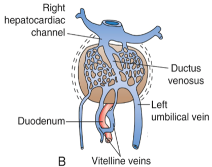
- Vitelline Veins
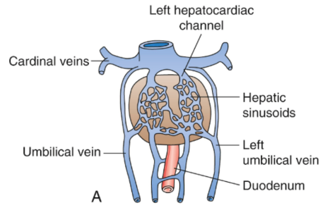
Timeline: 5 weeks
- Hepatic sinusoids have developed
- Vitelline (from gut) and umbilical veins (from mother) are beginning to drain into them
- Although there are channels that bypass these sinusoids
Second month
- Vitelline Veins (from gut) drain directly into the hepatic sinusoids
- Ductus Venosus has formed and accepts oxygenated blood from the Left Umbilical Vein
- Bypasses the hepatic sinusoids
- Directly enters the hepatocardiac channel
- Right umbilical vein begins to shrink
Third month
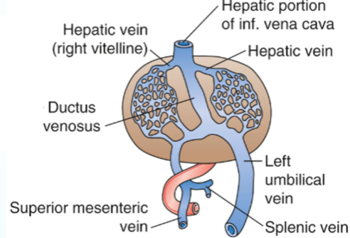
- Vitelline Veins have formed into the Portal system
- Splenic Vein
- SMV
- Portal Vein
- Right Umbilical Vein disappeared
- Left Umbilical Vein (future Ligamentum Teres) drains into the Ductus Venosus
- Then into Sinus Venosus/hepatocardiac channel
- Bypassing the hepatic sinusoids
- Note development of IVC + hepatic veins
- Overview of Fetal Circulation
- Fetal circulation supports the fetus by supplying oxygen and nutrients from the placenta.
- Blood flow is diverted away from the lungs and liver via shunts since these organs are not functional before birth.
- Key Structures in Fetal Circulation
- Placenta: Organ responsible for nutrient and oxygen exchange between the mother and fetus.
- Umbilical Cord:
- Contains two umbilical arteries carrying deoxygenated blood from the fetus to the placenta.
- Contains one umbilical vein carrying oxygenated blood from the placenta to the fetus.