Section: Endocrine Sub-section: Parathyroid
Calcium Transport
- Transported in the blood
- Bound to plasma proteins (45%; largely Albumin)
- Bound to small anions such as phosphate or citrate (15%)
- Free ionized state (40%)
- Normal range of ionized Ca2+ is 4.6 to 5.2 mg/dL
- Total serum Ca2+ concentrations range from 8.5 to 10.2 mg/dL
- Calcium levels are maintained within the very narrow range required for optimal activity of intracellular and extracellular processes
- Hormone secretion
- Muscle contraction
- Synaptic/Nerve function
- Coagulation cascade
- If fluctuation of protein concentration, total calcium levels vary significantly, but ionized calcium remains relatively stable
- E.g. volume overload, chronic illness, or malnutrition, serum protein is often reduced
- Low serum protein leads to a low total plasma calcium level; however, ionized calcium remains within normal limits
- Pseudohypocalcemia
- Changes in pH alter the equilibrium between calcium and albumin
- Acidosis reduces calcium binding to albumin
- Alkalosis enhances binding and can cause symptoms
- When significant shifts in pH, measure ionized calcium for accurate assessment of calcium status
Calcium Homeostasis
- Main regulators are
- PTH
- Vitamin D
- Calcitonin
- Main target organs of these are
- Bone
- Kidneys
- Gut
Parathyroid Hormone
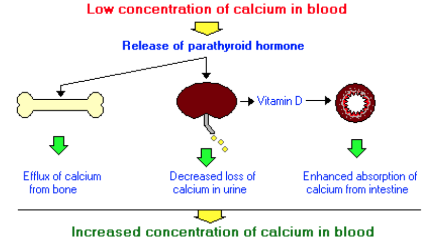
- Secreted by Parathyroid Chief Cells in response to decreased serum (extracellular) Ca2+
- Plasma ½ life of 4.5 mins
- Exerts action on target organs by binding to:
- PTH1R (Parathyroid 1 receptor)
- Heavily expressed in bone (osteoblasts) and kidney
- Also present in breast, skin, heart and pancreas
- Recognises PTH and PTHrP
- PTH2R (Parathyroid 2 receptor)
- Heavily expressed in GIT, CVS and CNS (rarely present on bone and kidneys)
- Selectively binds PTH only
- PTH1R (Parathyroid 1 receptor)
PTH Effects
- Kidneys
- Increased Ca2+ resorption from distal tubule
- Increased Phosphate and Bicarb excretion
- Increased renal synthesis of Calcitriol: 1,25-dihydroxyvitamin D3
- Stimulates synthesis of 1α-Hydroxylase in Proximal tubules
- Results in increased GIT Ca2+ and Phosphate absorption
- Bone
- Works in 2 phases
- Early, mobilises readily available Ca2+ stores - osteoblasts
- Later, indirectly stimulates bone resorption (Ca2+ and Phosphate release) - osteoclasts
- PTH1R on osteoblasts
- No PTH receptor on osteoclasts, but osteoclasts indirectly stimulated as action coupled to osteoblasts (by RANK ligand)
- Intermittent PTH stimulation causes bone deposition, chronic high PTH stimulation causes bone resorption
- Works in 2 phases
- Net effect
- Increases Serum Ca2+ and decreases Phosphate (if kidneys functioning)
Vitamin D
- Overview
- Initially in inactive forms
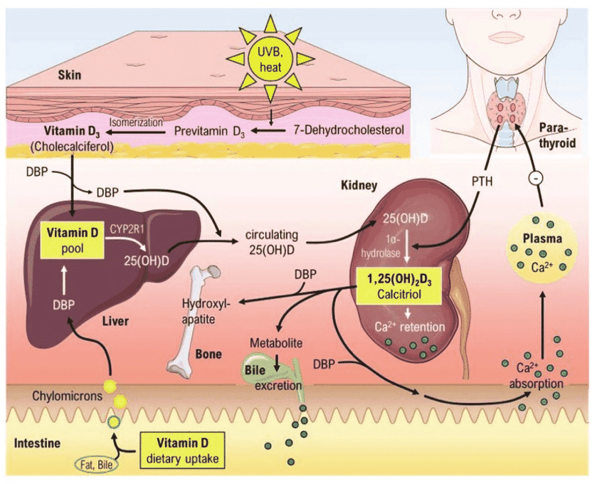
- Ergocalciferol (Vit D2) - from diet
- Cholecalciferol (Vit D3) - from sunlight
- Converted to Calcidiol (25-hydoxyvitamin D3) by hepatic enzyme 25-hydroxylase
- Calcidiol travels in circulation to kidneys and converted to Calcitriol (1,25-hydroxyvitamin D) which is the active form by 1α-Hydroxylase
- Initially in inactive forms
- Absorption/Synthesis
- Fat soluble vitamin (makes steroid hormone)
- Can have deficiency if fat malabsorption syndrome
- Most synthesised by UV light at the skin
- Small amount from diet
- Very few foods naturally contain Vit D
- Except fatty fish livers
- Most dietary Vit D from fortified foods and supplements
- Very few foods naturally contain Vit D
- Initially in inactive forms
- Ergocalciferol (Vit D2) - from diet
- Cholecalciferol (Vit D3) - from sunlight
- Fat soluble vitamin (makes steroid hormone)
- Calcidiol (25-hydoxyvitamin D3)
- Vit D2 and Vit D3 from gut and skin circulate to liver
- Hepatic enzyme 25-hydroxylase places hydroxyl group at 25 position of Vit D molecule
- Makes 25-hydroxyvitamin D (Calcidiol)
- Main circulating form of Vit D
- T1/2 is 2-3 weeks
- Has mild activity at bone and intestine
- < 1% as potent as Calcitriol
- Makes 25-hydroxyvitamin D (Calcidiol)
- Calcitriol (1,25-hydroxyvitamin D)
- Calcidiol travels in circulation to kidneys
- Taken up into Proximal Tubule cells
- 1α-Hydroxylase converts Calcidiol into 1,25-hydroxyvitamin D (Calcitriol)
- Most active form of Vit D
- T1/2 ~ 4-6 hours
- Calcitriol effects
- Most important: Promotes enterocyte differentiation and intestinal absorption of Ca2+
- Also stimulates intestinal phosphate absorption
- Suppression of PTH (negative feedback)
- Regulation of osteoblast function
- Permissively allowing PTH-induced osteoclast activation and bone resorption
- Decreased renal excretion of Ca2+ and phosphate
- Calcidiol travels in circulation to kidneys
- 1α-Hydroxylase
- Converts 25-hydroxyvitamin D to 1,25-dihydroxyvitamin D
- Mostly in the Proximal Tubule cells of the kidney
- Also present at extra-renal sites
- GIT, skin, vasculature, mammary epithelial cells, osteoblasts, and osteoclasts
- Most commonly seen extra-renal calcitriol synthesis is in granulomatous disease (e.g. Sarcoid)
- Get PTH independent extra-renal calcitriol synthesis
- Results in hypercalcaemia and hypercalciuria
- Regulated by
- Increased PTH, due to low Ca2+ (stimulates)
- Low phosphate (stimulate)
- FGF-23 (inhibits)
- Converts 25-hydroxyvitamin D to 1,25-dihydroxyvitamin D
Calcitonin
- 32 amino acid polypeptide secreted by parafollicular C cells
- Located superolaterally in each thyroid lobe
- Increased peripheral levels of Ca2+ stimulate calcitonin release
- Ca2+ infusion, gastrin/pentagastrin, ETOH can do this
- Major effect (“tones down calcium”)
- Acts on the surface receptors of osteoclasts
- Directly inhibits Ca2+ release from bone
- Decreases peripheral serum Ca2+ levels
- Minor Effect
- Inhibits renal tubular reabsorption of Ca2+ and phosphate
- Allows them to be excreted in the urine
- In normal physiology, calcitonin has minor role
- E.g. Medullary Thyroid Ca has calcitonin excess but has little alteration in peripheral Ca2+ metabolism
- E.g. Post-thyroidectomy: Low calcitonin doesn’t require Ca2+ supplementation
- Sensitive markers from primary or recurrent Medullary Thyroid Ca