Sub-section: Thyroid Section: Endocrine
Definition
- Thyroid follicular epithelial-derived cancer
- Subdivided into
General Principles
- Treatment options
- Conservative management
- Surgery
- TSH suppression
- Radioactive iodine treatment
- Similar principles for persistent/recurrent disease
- Radiation and systemic chemo play a role in a few cases
- Future therapies of targets tyrosine kinase inhibitors may be useful for metastatic DTC that is refractory to RAI
- Principles
- Remove primary tumour and clinically significant cervical nodes
- Minimise treatment related morbidity
- Accurately stage the disease
- Facilitate post-op RAI therapy if appropriate
- Permit accurate long-term surveillance
- Minimise risk of recurrence or metastasis
Conservative management
- For small papillary microcarcinoma, with no clinically evident metastasis, in an easily accessible area – you can consider active surveillance.
- This is useful for patients who’s co-morbidities confer a higher surgical risk, or who short remaining life-span.
- Contra-indications to conservative management include previous head and neck radiation, familial thyroid cancer syndrome, or evidence of nodal metastasis.
Thyroid Resection
Upfront strategy
- Thyroid Resection - PTC
- Consider Hemithyroidectomy if:
- < 1cm (some debate could be up to 4cm)
- Well-differentiated
- Unifocal
- No local invasion
- Total Thyroidectomy if any of
-
4cm
- High risk patients - Previous head/neck radiation/ Familial PTC
- Suspicious contralateral lesions
- Evidence of cervical LN mets
- Multifocal Papillary Microcarcinoma
- Definition: Size < 1cm
- < 5 foci – Hemithyroidectomy
-
5 foci – Total Thyroidectomy Size
- Definition: Size < 1cm
-
- <1cm (T1a)
- Hemithyroidectomy or conservative management.
- 1-4cm, without extra-nodal extension and lymph node negative (T1b – T2, No)
- Either a hemithyroidectomy or total thyroidectomy
- Factors which would make you do a total thyroidectomy vs lobectomy
- Need to consider the likelihood of there being residual disease in the other lobe i.e. other suspicious nodules.
- What you think the likelihood of needing radioactive iodine is (you can’t give radioactive iodine if there is a residual hemi-thyroid)
- Tumours >4cm in size
- Total thyroidectomy NB: it is uncommon to have a tumour >4cm without lymph node involvement.
- Any tumour size with extrathyroidal extension (T3+), lymph node metastasis, or distant metastasis
- Total thyroidectomy
- High risk patients (Previous head/neck radiation and Familial PTC
- Total thyroidectomy
Indications for subsequent completion thyroidectomy
- If initially thought to be well differentiated cancer
- Poorly differentiated high grade carcinoma
- Lymphatic or vascular invasion.
- Multifocal disease
- If Bethesda III or IV lesion and no indication for Total Thyroidectomy
- Completion Total Thyroidectomy if confirmed on histology
- Except if < 1cm (or maybe <4cm), unifocal, minimally invasive, intrathyroid, node neg
Follicular thyroid cancer surgical treatment
- You often won’t know it’s a follicular cancer pre-operatively.
- You will often be operating on a FNA results reporting a “follicular lesion of uncertain significance” (Bethesda III or IV lesion)
- You will often be doing a diagnostic lobectomy
- The management of T1-T2 tumours is the same as papillary thyroid cancer.
- <1cm leave
- 1-4cm discuss
-
4cm completion thyroidectomy
Lymph Node Dissection
Therapeutic LN dissection
- Do if exam or USS evidence of pathologic nodes (central or lateral)
- Therapeutic central-compartment (level VI) neck dissection for patients with clinically involved central nodes should accompany total thyroidectomy to provide clearance of disease from the central neck.
- Therapeutic lateral neck compartmental lymph node dissection should be performed for patients with biopsy-proven metastatic lateral cervical lymphadenopathy
Prophylactic LN dissection - PTC only
- Consider central (level VI) dissection if
- Tumour > 4cm +/- extrathyroidal extension (T3 or T4)
- Clinically involved lateral nodes
- High risk patients (Previous head/neck radiation, Familial PTC)
- Arguments
- For prophylactic central neck dissection
- Low morbidity if done in the hands of an experienced surgeon
- Gives more accurate staging (thus helps determine need for radioactive iodine).
- May reduce the need for re-operative surgery – there is a trial that looked at tumours >1cm, and found that there was 4x lower risk of re-operative surgery for recurrence in patients who had a routine central neck dissection
- Might lower post-op thyroglobulin levels which means it is more useful for monitoring.
- Against
- Most metastasis are micro-metastatic
- Removal of these occult foci does not clearly influence survival and they would likely be treated by radioactive iodine.
- Most metastasis are micro-metastatic
- For prophylactic central neck dissection
- Prophylactic lateral neck dissection not recommended
Radioactive Iodine Therapy (I131)
Overview
- Indications/Purposes
- Ablation of remnant thyroid tissue to facilitate detection of later recurrence by imaging and Tg
- Adjuvant therapy targeting occult metastatic disease
- Primary treatment of known persistent disease
- Not effective for Hurthle cell differentiated follicular cancer – only about 15% of these tumours will take up iodine.
- Pre-treatment considerations
- Need low iodine stores
- 1-2/52 low iodine diet to make cells iodine avid
- Avoid iodine loads
- Amiodarone – can interfere with RAI therapy for months to years
- Contrast CT has high load - can mean delay to therapy for up to 3 months
- 1-2/52 low iodine diet to make cells iodine avid
- Need high TSH levels (stimulates RAI uptake)
- Aim for levels > 30IU/L
- Can withdraw thyroid hormone
- W/H levothyroxine for 3-4/52 prior
- Can also give thyrogen, recombinant human TSH (rhTSH)
- Avoids hypothyroidism
- Using rTSH/thyrogen is easier for the patient - it can be given just before the RAI
- Evidence early but caution with intermediate/high risk Ca’s (can stimulate mets etc)
- Avoids hypothyroidism
- Need low iodine stores
Indications
- Indicated for differentiated thyroid cancers (papillary and follicular)
- Three reasons
- Remnant ablation
- Adjuvant treatment
- Treatment of known disease
- Based on risk
- High risk - give
- Intermediate risk - consider
- Low risk - do not give
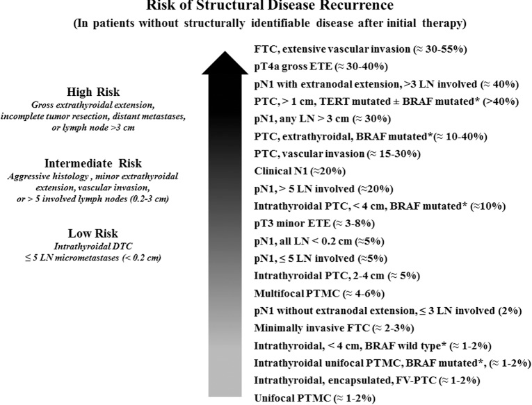
Summary of ATA risk
- Low risk – Papillary thyroid cancer confined to thyroid
- Intermediate risk – Low regional mеtаѕtaѕes, worrisome histologies, extrathyroidal extension, or vascular invasion
- High risk – High regional mеtаѕtaѕes, Gross extrathyroidal extension, distant mеtаѕtases, or postoperative serum Tg suggestive of distant mеtаѕtases
Low risk tumour
- Tumour <4cm
- No vascular invasion
- Not high grade
- Not a worrisome subtype.
- <5 lymph nodes which are micro metastatic (<2mm).
Intermediate risk tumour
- Tumour > 4cm
- Microscopic invasion into peri-thyroidal tissue
- Tumour with aggressive histology - tall cell, insular, Hurthle cell carcinoma etc.
- Clinical N1 or >5 pathologic N1 with all involved lymph nodes <3 cm in largest dimension
High risk tumour
- Risk
- Gross extra-thyroidal extension
- Pathologic N1 with any metastatic lymph node ≥3 cm in largest dimension
- Multiple lymph nodes >3cm
- Follicular thyroid cancer with extensive vascular invasion (>4 foci)
- Remnant disease
- R1
- Any evidence of distant metastatic disease.
- Thyroglobulin level suggestive of metastatic disease.
- High risk disease:
- Metastatic disease (M1)
- Gross extrathyroidal extension (T4)
- Consider if intermediate risk (and generally favoured in):
- Tumour > 4cm (T3)
- Microscopic extrathyroidal extension or
- Positive central (N1a), lateral or mediastinal LNs (N1b)
- Adverse histological subtype (Tall cell) or vascular invasion
- Not routinely recommended if low risk
- 1 - 4cm, intrathyroidal, no nodes or mets
AMES risk calculator
- AMES
- Age
- Metastases
- Extrathyroidal invasion
- Size
- Gives low risk or high risk
Dose
Remnant ablation
- Is typically 30mCi
- Primary goal is destruction of remnant thyroidal tissue to facilitate staging and follow up studies.
- There is controversy regarding the role of remnant ablation - this is something which is generally only offered to patients with low risk tumours, which, generally don’t benefit from radioactive iodine treatment anyway.
Adjuvant therapy
- Is typically 75mCi
- to treat any suspected but not proven disease (in moderate or high risk patients).
Therapeutic
- Is typically 150mCi
- patients with known residual or recurrent disease (biochemically or structurally)
Contraindications
- Pregnancy – Crosses placenta and causes Cretinism
- Breast-feeding
- Lactating breast will take up radioiodine
- Need to stop feeding 6-8/52 prior to Rx (or consider dopamine agonist at later time)
Complications
- Sialadenitis
- Neck oedema
- Tumour haemorrhage/oedema
- Risk of secondary malignancies
- Salivary or breast (have Na/I symporter)
- Urinary or GIT (RAI excreted from here)
- Reduce by giving laxatives and encourage hydration to clear RAI
- Leukaemia
- Myelodysplastic syndromes
- Need to advise to stay 6 feet away from others for 24hrs
Follow up
- Post RAI imaging should occur 2-8 days after RAI is given.
- You will be able to observe WHERE the RAI has been taken up - which will thus confirm metastatic disease sites and thus help with staging.
- If a metastatic site doesn’t take up radio-iodide on this scan - then it is not responsive to RAI and treatment should not be repeated.
TSH Suppression
- Differentiated Thyroid Ca express TSH-R
- TSH still works as growth factor
- Give supra-physiologic dose of Thyroxine
- Balance between risk of malignancy and hyperthyroid symptoms
- Aim
- High Risk
- Aim TSH < 0.1 mU/L
- Intermediate Risk
- Aim TSH 0.1-0.5 mU/L
- Low Risk
- No suppression
- Aim TSH 0.5-2.0 mU/L
- High Risk
- Long term consequences of TSH suppression
- Arrhythmias/AF
- Development of osteopenia/osteoporosis
Follow up
Thyroglobulin
- Check serum Tg levels 4-6 weeks post-op
- Normal should be:
- Total thyroidectomy + ablation
- Nonstimulated Tg <0.2 ng/mL
- TSH-stimulated Tg <1 ng/mL
- Total thyroidectomy
- Nonstimulated Tg <0.2 ng/mL
- TSH-stimulated Tg <2 ng/mL
- Lobectomy
- Serum Tg level <30 ng/mL
- Total thyroidectomy + ablation
- Stimulating vs non-stimulation
- Most patients will be on thyroxine (TSH suppressed)
- Patients can be given rhTSH or thyroxine stopped (TSH stimulated) to increase the level of Tg
- Stimulated Tg values are useful to document an excellent response to therapy or to identify the presence of persistent/recurrent disease
- With the newer, more sensitive Tg assays stimulation may not be required
Dynamic risk stratification
- Reassessment of response to therapy at each visit
- Based on
- Neck USS
- TSH
- Thyroglobulin
- Based on
- Classifications
- Excellent response to treatment
- Negative imaging (USS or radio-iodide scan)
- Non-stimulated thyroglobulin < 0.2
- Stimulated thyroglobulin level < 1.
- Indeterminate response
- Non-specific signs on imaging (USS or radio-iodide scan)
- Non-stimulated thyroglobulin level which is detectable but < 1
- Stimulated thyroglobulin level which is detectable but < 10.
- Biochemical incomplete response
- Negative imaging
- Non-stimulated thyroglobulin level > 1
- Stimulated thyroglobulin level > 10.
- Rising anti-thyroglobulin anti-bodies after treatment.
- Structurally incomplete response
- Residual disease on USS or radio-iodide scan
- With or without elevated thyroglobulin levels.
- Excellent response to treatment
Surveillance
- Usually follow up appointment to check wound
- Check TFT and baseline thyroglobulin at 6-8 weeks post-op
- Neck USS
- 6–12-month intervals
- TSH + Thyroglobulin
- Every 3-6 months
- NB: Also measure anti-Tg levels as antibody will interfere with assay
- If concerns on USS or biochemically, perform
- Diagnostic whole body radioiodine scan
- Consider CT, MRI, PET-CT
Recurrence
- Minimal disease
- Re-operative an option
- If on going uptake on radioiodine scanning then RAI can be given
- Disease that is FDG-PET avid tends to not respond to RAI
- Extensive disease
- Radioiodine, if scans demonstrate uptake.
- Systemic chemotherapy (eg, kinase inhibitors).
- External radiotherapy.
- Percutaneous ethanol injection of cervical nodal metastases.
- Radiofrequency ablation of cervical, osseous, and pulmonary metastases – This is an alternative for patients who are poor surgical candidates and whose metastases do not concentrate radioiodine, but expertise in this treatment modality is not widely available.
- Palliative embolization of bone metastases – Palliative embolization may reduce symptoms or be used prior to surgery.