Sub-section: Thyroid Section: Endocrine
Definition
- Originates in Parafollicular cells (C cells)
- Upper /middle areas of the thyroid lobes
- Neural crest origin – 4th and 5th Branchial Pouch
- Join the thyroid during development
- C-cells produce calcitonin in the thyroid
Incidence
- 4% to 10% of Thyroid Carcinomas
- 13% of Thyroid Ca deaths
Classification
- Sporadic (75%)
- Hereditary (25)
- MEN 2A syndrome
- Medullary Thyroid Ca
- Phaeochromocytoma
- Parathyroid Hyperplasia
- MEN 2B syndrome
- Medullary Thyroid Ca
- Phaeochromocytoma
- Hirschsprung’s Disease
- Mucosal Neuroma
- Familial Medullary Thyroid Carcinoma (FMTC)
- Variant of MEN2A that includes MTC but not the other features of MEN2A
- MTC arising in MEN2A usually has more favourable long-term outcome than MEN2B or sporadic
- MEN 2A syndrome
Clinical Features
- A single palpable mass, usually painless (most cases)
- Elevated calcitonin
- Systemic symptoms may occur due to hormonal secretion by the tumour - which includes diarrhoea and facial flushing
- In most patients, there is metastasize at diagnosis - usually to nodes.
Genetics
- MEN2 and FMTC involve different germ line activating mutations in the RET proto-oncogene
- NB: Also 40-50% sporadic MTCs have acquired RET mutations
- Inherited MTC’s develop C-Cell Hyperplasia
- Pre-neoplastic lesion if RET mutation
- No/little malignant potential if no RET mutation
- Pre-neoplastic lesion if RET mutation
- MTC has high penetrance (> 90%)
- Family members of patients with MEN2 should be screened at an early age for the RET proto-oncogene.
- Shortly after birth in MEN2B kindreds
- Before age 5 years in FMTC and MEN2A kindreds
- Work up when any patients is diagnosed with MTC
- History
- Workup includes a detailed and in-depth family history
- Cancer history
- Inquire about the characteristics of MEN2 in the patient and family members
- Workup includes a detailed and in-depth family history
- Parathyroid
- Serum Ca2+ and PTH
- Phaeochromocytoma
- Urinary Catecholamines (Phaeochromocytoma)
- Must be excluded before considering interventions in patients with MTC
- Refer to genetics for RET testing
- History
Investigations
- Serum Calcitonin
- If the pre-operative calcitonin is >500pg/mL - CT neck CAP is required.
- CEA
- USS + FNA
- USS must include evaluation of the neck nodes
- Refer to genetics
- Genetic testing for germline RET mutation
- Biochemical evaluation for Phaeochromocytoma and parathyroid
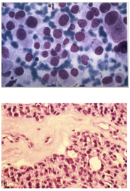
Histopathology
- FNA
- Round blue cells
- Increased Amyloid deposition (Stains with Congo Red)
- Histology
- Arise from calcitonin cells - thus is a form of neuroendocrine tumour.
- The histopathological appearance can be quite variable - thus staining is very important
- Stain positive for calcitonin, CEA, chromogranin, and synaptophysin.
- Will not stain for thyroglobulin (as not from follicular cells)
Biochemical testing
- Calcitonin
- Shown to be an effective marker for the presence of MTC
- NB: Elevated calcitonin in absence of a thyroid mass requires further workup
- Repeat calcitonin
- Calcium-stimulated or gastrin-stimulated test
- NB: Elevated calcitonin in absence of a thyroid mass requires further workup
- Calcitonin excess is not clinically associated with hypocalcaemia
- May rarely result in symptoms of diarrhoea and flushing in advanced disease
- Calcitonin levels represent volume of disease
- Presence of a mass and an elevated calcitonin level is virtually diagnostic of MTC
- Shown to be an effective marker for the presence of MTC
- CEA
- CEA may also be elevated in MTC
Grading
- Because it is a neuroendocrine tumour - can be defined as low grade or high grade based on Ki67, mitosis for 2mm2, and evidence of tumour necrosis.
- Low grade - <5 mitosis/10HPF, Ki67 <5 %, No tumour necrosis.
- High grade - >5 mitosis/10HPF, Ki67 >5 %, tumour necrosis
Management
- Local disease
- Total Thyroidectomy with Prophylactic Level VI Central Nodal Dissection
- Lateral neck dissection if positive lymph nodes
- Lateral neck dissection based on Calcitonin
- Controversial
- If the calcitonin level is >200pg/mL - ipsilateral lateral lymph node dissection.
- Bilateral neck dissection unlikely to confer benefit
- If micomets here then likely to be elsewhere
- Controversial
- Known nodal metastasis in the central neck
- Level VI Central Nodal Dissection
- Some advocate the addition of a lateral neck dissection on the side ipsilateral to the level VI disease
- (not universally recommended)
- Clinically detectable or ultrasound-detectable disease in the lateral neck
- Total Thyroidectomy, Level VI node dissection and ipsilateral lateral compartment nodal dissection
- Some people advocate for contralateral lateral neck dissection if calcitonin level is > 200pg/mL
- Distant metastatic disease
- Less aggressive surgery in the neck to decrease the risk of morbidity
- Palliative operations may be indicated in patients with neck pain or airway compromise
- Other options to treat metastatic disease
- Radiotherapy to bony metastasis
- Embolisation
- Tyrosine kinase inhibitors
- Palliative cytotoxic chemotherapy - 5FU etc
- If MTC is diagnosed post-op in a pt undergoing less than Total Thyroidectomy
- Further surgery to complete therapy as though known pre-op
- Completion Thyroidectomy and Nodal Dissection as indicated
- Exceptions: Incidental finding of MTC in thyroid lobectomy where:
- MTC is sporadic and unifocal
- No C-Cell Hyperplasia
- Otherwise normal USS neck
- Negative surgical margin
- Normal serum calcitonin
Prophylactic Thyroidectomy
- MEN2B RET mutation
- Within the 1st year or at the time of dx
- Other patients with germline RET mutations
- Before age 5 years or at the time of dx
- May be appropriate to wait beyond 5 years with particular RET mutations
- Don’t need to do level VI node dissection in above patients unless
- Thyroid nodules > 5 mm
- Elevated calcitonin
- Lymph node metastasis
Follow-up
- Calcitonin and CEA
- Basal and stimulated calcitonin levels to monitor for recurrence
- Calcitonin values may increase before basal calcitonin levels
- Can do stimulated calcitonin test but has minimal clinical utility
- Calcitonin values may increase before basal calcitonin levels
- Basal and stimulated calcitonin levels to monitor for recurrence
- Recurrent MTC found biochemically is often associated with unresectable recurrence in distant metastatic locations
- E.g. Lung and liver
- TSH suppression and RAI scanning and therapy have no role in MTC
- Unless there is a concomitant PTC or FTC