Sub-section: Thyroid Section: Endocrine
Definition
- The thyroid gland is an endocrine organ in the central neck that predominant function is to create thyroid hormone T3 and T4
Functions
- Collect and transport iodine
- Synthesises Thyroglobulin and secrete it into colloid
- Removal of T4 and T3 from Thyroglobulin and secretion into circulation
- Produces 3 Hormones:
- Thyroxine (T4)
- Triiodothyronine (T3)
- Calcitonin
- Functions of Thyroid Hormones
- Stimulate O2 consumption of most cells
- Help regulate lipid and CHOH metabolism
- Necessary for normal growth and maturation
- Thyroglobulin
- Glycoprotein that facilitates MIT + DIT → T3 + T4
- Process accompanied by release of thyroglobulin into blood (can be measured)
Iodine
- Iodine essential for production of thyroid hormones
- Dairy, eggs, iodised salt, saltwater fish, shellfish, soy, multivitamins
- Absorbed via GIT as inorganic iodide and enters extracellular iodide pool
- 90% body iodide stored in Thyroid
- Stored as pre-formed thyroid hormone
- Transported from extracellular space into Follicular cells
- Against chemical and electrical gradient at baso-lateral membrane by Na+-I symporter (NIS)
- Regulated by TSH from Pituitary Glands as well as follicular iodide content
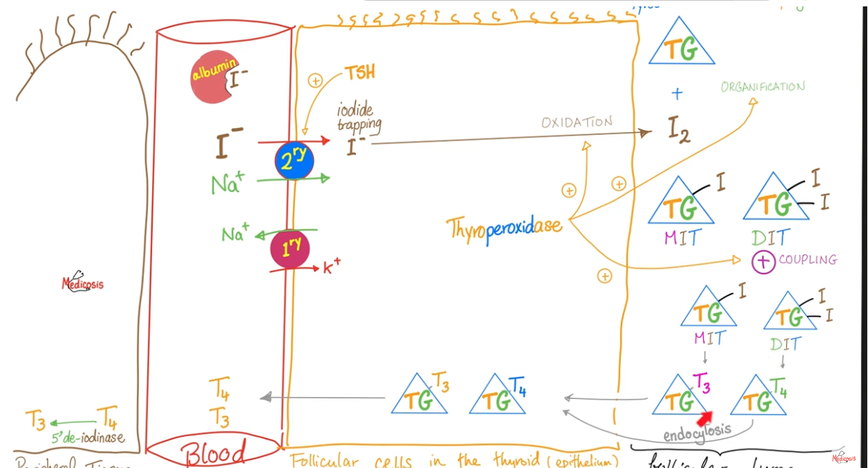
Thyroid Hormone Synthesis
- Synthesis of Thyroglobulin: Thyrocytes in the thyroid follicles produce a protein called thyroglobulin (TG).
- TG does not contain any iodine, and it is a precursor protein stored in the lumen of follicles. It is produced in the rough endoplasmic reticulum.
- Iodide uptake: Protein kinase A phosphorylation causes increased activity of basolateral Na+-I- symporters, driven by Na+-K+-ATPase, to bring iodide from the circulation into the thyrocytes. Iodide then diffuses from the basolateral side to the apex of the cell, where it is transported into the colloid through the pendrin transporter.
- Iodination of thyroglobulin: Protein kinase A also phosphorylates and activates the enzyme thyroid peroxidase (TPO). TPO has three functions: oxidation, organification, and coupling reaction.
- Oxidation: TPO uses hydrogen peroxide to oxidize iodide (I-) to iodine (I2).
- Organification: TPO links thyroglobulin protein with iodine. It generates monoiodotyrosine (MIT) and diiodotyrosine (DIT). MIT has a tyrosine residue with iodine, and DIT has tyrosine residues with 2 molecules of iodine.
- Coupling reaction: TPO combines iodinated tyrosine residues to make triiodothyronine (T3) and tetraiodothyronine (T4). MIT and DIT join to form T3, and two DIT molecules form T4.
- Storage: thyroid hormones are bound to thyroglobulin for storage in the follicular lumen.
- Release: thyroid hormones are released into the fenestrated capillary network by thyrocytes in the following steps:
- Thyrocytes uptake iodinated thyroglobulin via endocytosis
- Lysosome fuse with the endosome containing iodinated thyroglobulin
- Proteolytic enzymes in the endolysosome cleave thyroglobulin into MIT, DIT, T3, and T4
- T3 (20%) and T4 (80%) are released into the fenestrated capillaries via MCT8 transporter.
- Deiodinase enzymes remove iodine molecules from DIT and MIT. Iodine can be salvaged and redistributed to an intracellular iodide pool.
Thyroid Peroxidase (TPO)
- Enzyme that has central role in thyroid hormone synthesis
- Actions
- Oxidises Iodide to iodine
- Organification (binding of iodine to tyrosine residues of thyroglobulin)
- Coupling of MIT/DIT to T3/T4
- Actions
- Stimulated by TSH:
- Upregulates gene expression
- Inhibited by Thioamides
- Hashimoto thyroiditis has (among others) anti-TPO antibodies that inhibit the function of TPO
Thyroglobulin (Tg)
- 660-kDa glycoprotein specific to follicular cell
- Primary component of colloid matrix necessary for iodination
- Facilitates conversion of MIT and DIT to T3 and T4
- Process leaks small amounts of Tg into blood where it can be measured
- Excess peripheral levels of iodine inhibit further release by enhancing Tg resistance to proteolysis
- Peripheral Tg can be measured to evaluate benign or malignant neoplasms
- Predictive value for measuring local or metastatic recurrence post-thyroidectomy in Differentiated Thyroid Ca
- Usefulness of pre-op measurement not-known, not recommended
Calcitonin
- 32 amino acid polypeptide secreted by Parafollicular C cells
- Located superolaterally in each Thyroid lobe
- Acts on the surface receptors of osteoclasts
- Inhibits Ca2+ release from bone
- Decreases peripheral serum Ca2+ levels
- Increased peripheral levels of Ca2+ stimulate Calcitonin release
- Ca2+ infusion, Gastrin/pentagastrin, EtOH can do this
- In normal physiology, Calcitonin has minor role
- E.g. Medullary Thyroid Ca has Calcitonin excess, but has little alteration in peripheral Ca2+ metabolism
- E.g. Post-thyroidectomy, low Calcitonin doesn’t require Ca2+ supplementation
- Basal or stimulated Calcitonin levels are sensitive markers from primary or recurrent Medullary Thyroid Ca
Regulation of Thyroid Hormone Secretion
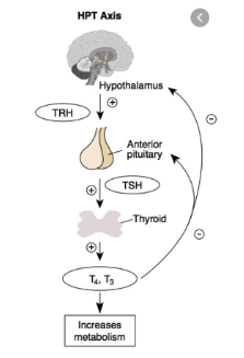
- Hypothalamic-pituitary-thyroid axis
- Major regulator is TSH, which stimulates
- Thyroid cell growth and differentiation
- Iodine uptake
- Tg synthesis
- Release of T3 and T4 from Tg
- Also shown to stimulate growth and invasive characteristics of differentiate thyroid cancers
- TSH secreted in pulsatile fashion from Anterior Pituitary
- Binds to TSH receptor on Thyroid Cells (TSH-R)
- Stimulates G-protein and cAMP pathway
- Mutations in genetics of this system are associated with follicular neoplasms
- Feedback loop important
- Decreased T3 results in release of increased thyroid releasing hormone (TRH) from Paraventricular Nucleus of Hypothalamus
- Stimulates increased TSH release from Anterior Pituitary
- Similarly T3 directly inhibits release and synthesis of TSH (primary hyperthyroidism)
- NB: Also intrinsic thyroid auto-regulatory mechanisms
- If excessive iodide ingested, auto-regulation inhibits uptake of excess
- If iodide deficiency, uptake increased
Peripheral Actions of Thyroid Hormones
- T3 significantly more bioactive than T4
- T4 converted peripherally
- T3 has high affinity for thyroid hormone receptor (TR)
- Both > 99% carried by binding proteins
- 80% TBG, the rest Albumin and Pre-Albumin
- TR is a steroid receptor
- T3/TR complex binds to regulatory regions in various gene-related processes
- TR production regulated by 2 genes located on chromosomes 17 and 3
- CNS contains α - form
- Liver contains β – form
- Physiological effect of T3 (+T4) is evident in all metabolically active tissues
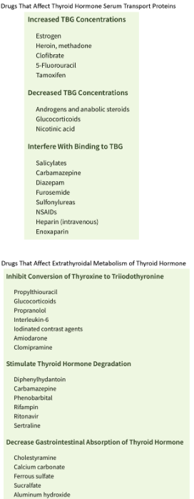
Drugs That Affect Thyroid Hormone Metabolism
Tests of Thyroid Function
- Evaluation of Pituitary-Thyroid Feedback Loop (TSH)
- Ultra-sensitive radioimmunoassay
- Important screening test for thyroid dysfunction
- Delineates hyper/hypothyroidism from euthyroid states
- Sensitivity means can detect dysfunction before clinical manifestations apparent
- Not affected by changes in binding proteins or non-thyroidal disease
- Free T3 and T4 levels
- Screening test of thyroid production levels
- NB: Assays for Total T3/4 affected by changes in hormone production or binding proteins
- Calcitonin
- Obtain baseline level if Thyroid mass and suspicion for MEN2 or Medullary Ca
- If doubt could do pentagastrin or Ca2+ stimulated levels
- Can use as screening test in families with MEN2
- Not recommended for routine use in workup of otherwise unsuspicious nodules
- Radioactive Iodine Uptake (RAI)
- Thyroid Autoantibodies
Radioactive Iodine (RAI) Uptake - Diagnostic I123
- Directly evaluates thyroid gland function
- Less widely used now due to more precise biochemical measurements
- I 123 given orally and uptake calculated with radioscintigraphy
- Normal result 15-30% uptake at 24hrs
- NB: Ablation uses I-131 which has longer ½ life and more radiation exposure
- Indications:
- If solitary nodule and low TSH (? Functioning hot nodule, ? Cause of hyperthyroidism)
- Detects functioning Thyroid Ca mets after remnant iodine ablation
Thyroid Autoantibodies
- Thyroid stimulating immunoglobulin
- Anti-microsomal antibodies
- Anti-thyroid peroxidase antibodies
- Produced in autoimmune thyroid disorders
- Grave’s Disease
- 80% have antimicrosomal antibodies
- Antibodies have high affinity for TSH-R on Follicular cells
- Assays have good sensitivity, allowing early detection and accurate monitoring
- Hashimoto thyroiditis
- 95% have detectable antimicrosomal antibodies
- TgAb highly sensitive marker of Hashimoto’s disease