Section: Endoscopy Curriculum: Curriculum, page 38
Resources:
- Cotton and Williams’ Practical Gastrointestinal Endoscopy: The Fundamentals, Seventh Edition
Endoscopic equipment
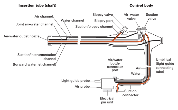
- Endoscope consists of:
- Control head with valves (buttons) for air insufflation, wash and suction
- Flexible shaft carrying light guide and service channels
- Bending section at tip
- Umbilical cord connecting endoscope to light source and processor, air supply, wash and suction.
- Biopsy channel – approx. 3 mm.
Ancillary equipment
- Suction traps: can be used to take samples of secretions
- Biteguards: to protect patient’s teeth and scope
- Overtubes: flexible plastic sleeves that cover the endoscope shaft and act as conduit for repeated intubations or facilitate therapeutic procedures such as extraction of foreign body and haemostasis
- Caps: attached to tip to facilitate banding and mucosal resection, dissection
- Stretchers/trolleys: Need to have side rails and ability to tilt head down
- Image documentation: video sequences are recorded digitally and can be printed
- Sedation and monitoring: require pulse oximetry as minimum. Usually have continuous BP monitoring and ECG. Appropriate resuscitation equipment available, including oral airways, oxygen delivery systems and wall suction. Endoscopic accessories:
- Biopsy forceps: consist of pair of sharpened cups, spiral metal cable, pull wire and control handle.
- Cytology brush: covering plastic sleeve to protect specimen during withdrawal
- Flexible needles: injections and sampling fluids
- Fluid-flushing devices: can do manual or electric pump flush of channel. Small syringe (1 – 5 ml) generates more pressure than a large one (50 ml) which generates more suction.
- Others: snares, clips
Argon plasma coagulation
- Uses electrical conductivity of argon gas- similar phenomenon to neon lights. Argon passed down electrode catheter and energised with an intelligent-circuitry electrosurgical unit and patient plate, ionises to produce a local plasma arc- like miniature lightning strike. Heating effect is superficial (2-3 mm), unless current applied in same place for many seconds, due to coagulation increasing resistance, causing plasma arc to jump elsewhere.
Reprocessing scope
- Scopes cannot tolerate heat > 60’C so cannot be sterilised (e.g. using autoclave), so must be reprocessed using high level disinfection. See Sterilisation and disinfection
- See https://www.youtube.com/watch?v=3B_SC_y2Kew
- See https://www.ncbi.nlm.nih.gov/pmc/articles/PMC4604272/#:~:text=Clean%20the%20external%20surface%20of,cleaned%20with%20an%20ultrasonic%20cleaner
- 7 steps:
- Precleaning: a. Wipe down with detergent solution b. Air and solution passed through channels repeatedly c. Transport in closed container
- Cleaning and brushing a. Leak test first b. Clean with low foam enzymatic detergent using brush and soft cloths
- Rinsing a. Flushing endoscope with sterile water
- High level disinfection: a. Immersed in disinfection solution - Glutaraldehyde. Can use automated endoscope reprocessor
- Final rinse with sterile water
- Dry channels – can use heated pressurised air.
- Storage in safe sterile environment, hanging in vertical position without touching floor.
Methods to improve detection of pathology
- NBI – optical filters to select wavelengths of light which correspond to the park light absorption of haemoglobin, enhancing visualisation of blood vessels and certain surface structures.
- Chromoendoscopy