Section: Hepatobiliary Sub-section: Pancreas Curriculum: Curriculum, page 90
Notes based on the 2024 Kyoto guidelines from the International association of Pancreatology (IAP) - International evidence-based Kyoto guidelines for the management of intraductal papillary mucinous neoplasm of the pancreas, page 1
Definition
- Grossly visible, mucin-producing epithelial neoplasm of the pancreas, which arise from within the main pancreatic duct or branch.
- Most often has papillary architecture.
- Distinguished from mucinous cystic neoplasms (MCNs) by absence of ovarian-type stroma
Three types:
- Main duct (MD-IPMN) - segmental or diffuse dilation of the MPD of >5 mm
- Branch duct (BD-IPMN) - cyst >5mm that communicates with the MD
- Branch type less likely associated with malignancy.
- Mixed type
Epidemiology
2.04 per 100,000, increases after 6th decade
Risk factors
Aetiology
- Aetiology unknown.
- Associated with extrapancreatic primaries (10%)
- Most commonly colorectal, breast and prostate.
- Predictor of pancreatic cancer (odds ratio 7.18)
Pathology
Microscopically
- Surrounding pancreatic parenchyma may appear firm and hard due to scarring and atrophy from obstructive chronic pancreatitis secondary to tumour.
- Typical appearance is mucin-secreting columnar epithelium with variable atypia
- Growth pattern varies from flat ducts (ectasia) through to prominent papillae
Morphological subtype
Three types
- Intestinal
- Gastric
- Most often low grade, most favourable prognosis
- Pancreatico-biliary type differentiation
- Highest risk of neoplastic progression
Grade
- IPMN microscopically demonstrates papillary growth of columnar neoplastic cells with mucin hypersecretion.
- Graded
- Low grade dysplasia (LGD)
- High grade dysplasia (HGD)
- invasive carcinoma (IC)
Genetic alterations
Frequent mutations
- KRAS (60–70 %)
- GNAS (50–70 %)
- RNF43 (15 %)
Location
- Location
- Head (70%)
- Diffusely (5-10%)
- Body or tail (20-25%)
Differential diagnsis
- IPMN vs PanIN (pancreatic intraepithelial neoplasia)
- IPMN – taller more complex papillae
- Abundant luminal mucin
- Cystic pancreatic endocrine neoplasm vs. IPMN
- Presence of coarse and stippled chromatin with a smooth nuclear membrane.
Clinical
Clinical presentation
- Symptoms related to pancreatic duct obstruction.
- 60 patients from John Hopkins group
- Abdo pain (59%)
- Jaundice (16%)
- Weight loss (29%)
- Diabetes
- 14% had previous acute pancreatitis
- 60 patients from John Hopkins group
- Incidental from imaging
Investigations
CT or MRI:
Classical features
- Main duct – segmental or diffuse main pancreatic duct dilatation >5mm.
- Branch duct – cystic lesions >5mm, grape like configuration
- Mixed – both of above
High risk stigmata for malignancy:
- Enhanced mural module or solid component >5mm
- Jaundice
- Main pancreatic duct size >10mm
- Positive or suspicious cytology
Worrisome features
- Acute pancreatitis
- Cyst size >3 cm
- Cystic growth rate >2.5mm/year
- Increased level of serum Ca19-9 (>37 U/mL)
- Thickened enhanced cyst walls
- Enhancing mural nodules <5 mm
- MPD size 5–9.9 mm
- Abrupt change in MPD calibre with distal pancreatic atrophy
- Lymphadenopathy
- New onset or acute exacerbation of diabetes within one year
Differential diagnosis
- Differentiating branch type IPMN:
- Difficult to differentiate from MCN or pseudocysts (need to consider patient’s age, gender, history of pancreatitis or genetic syndromes).
- Suggest IPMN
- Localisation within uncinate
- Detection of non-gravity-dependent luminal filling defects (papillary projections) or grouped gravity dependent luminal filling defects (mucin)
- Upstream dilatation of ducts (MCN ducts are normal)
- Differentiating diffuse main duct IPMN (from chronic pancreatitis):
- Patients with IPMN tend to be 20 years older, lack heavy EtOH hx
- On imaging
- Endoluminal filling defects (either mucin or papillary proliferations)
- Cystic dilatation of collateral branches (particularly uncinate process)
- Communication of dilated ducts with normal ducts without evidence of obstructing lesion
- Widely open papilla
EUS
- Advantage of being able to sample cystic fluid and biopsy solid lesions.
- Useful to detect high risk/worrisome features
Cyst fluid
- Purpose
- Discriminate mucinous cysts from other cysts
- Mucinous cysts - precursors for HGD/IC and thus require resection or surveillance)
- IPMN- intraductal papillary mucinous neoplasm
- IOPN - Intraductal oncocytic papillary neoplasm
- ITPN- intraductal tubulopapillary neoplasm
- MCN - Mucinous cystic neoplasm
- Other cysts
- Serous cystadenoma/serous cyst neoplasms
- Pseudocysts
- Mucinous cysts - precursors for HGD/IC and thus require resection or surveillance)
- Distinguish between LGD and HGD/IC.
- Discriminate mucinous cysts from other cysts
- Tumour markers within cyst fluid not proven to be accurate
- Molecular assessment is useful
- Elevate CEA suggests mucinous cyst (but can be elevate in psydocyst) and 30% of IPMN will have a low CEA
ERCP
- Can be used.
- Being replaced by MRI.
- Observation of mucin protruding from widely open papilla is diagnostic.
Tumor markers
- No tumour markers specific.
- Ca 19-9 (not CEA) shown to be independent predictor of malignancy.
Management algorithm/surveillance
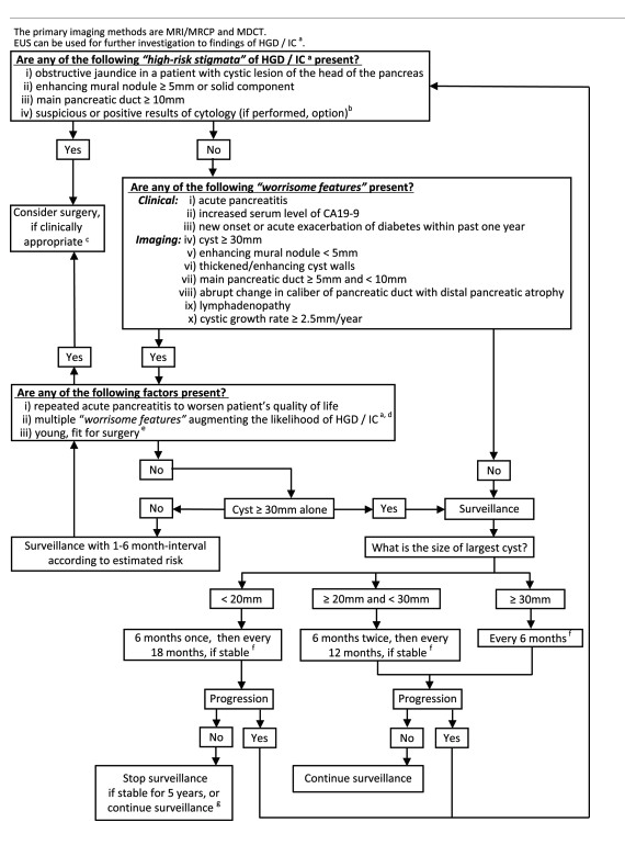 In a patient fit for surgery
- Operate on
- Any high risk features
- Multiple worrisome features
- Otherwise surveillance imaging every 6-12 months
- In some smaller IPMNs without change over 5 years they can be discharged from follow up.
Operative management
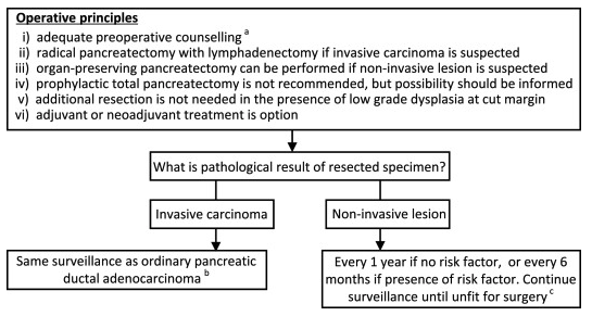
Suspicion for invasive carcinoma
- Radical pancreatectomy with lymph node dissection Low suspicion for invasive carcinoma
- Organ-preserving pancreatectomy without lymphadenectomy
- middle pancreatectomy or spleen-preserving distal pancreatectomy
Intra-operative considerations
- Intra-operative frozen section of the main pancreatic duct
- HGD or IC - additional resection
- LGD - additional resection not required
- May need to continue to total pancreatectomy
- In total pancreatectomy: long term risk of hypoglycaemia
Adjuvant treatment
- If IC found then as per pancreatic cancer
Outcome
- Main determinant of survival is presence of invasive disease
- Factors associated with poor survival:
- Presence of jaundice
- Tumour type (tubular worse than colloid)
- Vascular invasion
- Perineural invasion
- Poorly differentiated
- % tumour that was invasive
- Positive lymph nodes (41% of invasive disease)
- Margin status not associated with worse long-term outcome
Follow up
-
Role of adjuvant therapy for invasive IPMN not addressed in trials. In retrospective series, role is unclear
-
Recurrence
- Disseminated (arising from invasive disease)
- Local (within remnant, may or may not be invasive)
-
For invasive or dysplasia – MRI every 1-2 years
-
For those without above – no ongoing surveillance (except mixed-type IPMN or family history of pancreatic cancer)