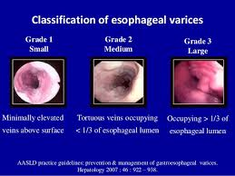
Oesophageal varicies classification
-
Low risk varices
- Small, minimally elevated with normal mucosa
-
High risk
- Small with red wale markings
- Medium (<1/3)
- Large (>1/3)
-
Grade
Primary prevention of variceal hemorrhage
- Screening
- Indication
- All patients with decompensated сirrhоsis
- Patients with compensated сirrhοsiѕ and either liver stiffness measurement (LЅМ) >20 kPa by transient elastography or platelet count <150,000/microL
- Indication
- Findings
- Low risk varices
- Screening in 1-2 years
- High risk
- NSBB
- In patients unable to take beta-blockers:
- Variceal banding
- This should be done every 2-4 weeks until eradication. After eradication an OGD should be performed every 3-6 months for the first year. Followed by screening every 2-3 years.
- Low risk varices
Acute management of variceal bleeding
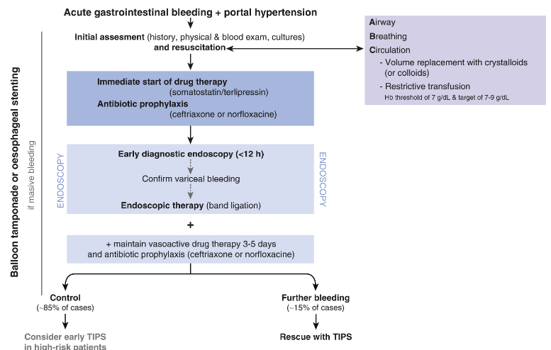
Pharmacological treatments
- Vasoactive medications (acute variceal hemorrhage)
- Cause splanchnic vasoconstriction
- Terlipressin (synthetic analogue of vasopressin)
- Preferred as has been shown to reduce mortality
- Somatostatin and Octreotide (somatostatin analog)
- Terlipressin (synthetic analogue of vasopressin)
- Cause splanchnic vasoconstriction
- Antibiotics (acute bleed)
- Gram -ve cover to reduce bacterial infections and decreased early re-bleeding
- PPI not indicated unless associated peptic ulcer disease
Endoscopy
- Can be used as primary prophylaxis if contraindication to beta blockers
- For oesophageal varices, endoscopic band ligation better than sclerotherapy (which is associated with higher rates of re-bleeding, mortality and stricture)
- For gastric varices, sclerotherapy with tissue adhesives such as cyanoacrylate more effective and safer than EBL
Balloon tamponade
- Temporary measure
- Intubate first
- Sengstaken-Blakemore tube
- Gastric balloon is inflated in the stomach (e.g. with 30 mL of water) and placed on gentle traction against the gastro-oesophageal junction
- Inflation of the oesophageal balloon should be delayed and only undertaken if bleeding persists as there is an increased risk of necrosis at the gastro-oesophageal junction
- Oesophageal balloon must be deflated every 12 hours to prevent necrosis, and for similar reasons the gastric balloon should not be inflated for more than 48 hours
- Complications – aspiration pneumonia, oesophageal rupture, asphyxia from balloon migration, oesophageal ulcers, pressure necrosis (tongue or lips), arrhythmia and chest pain
- Self-expandable metal stent may be a better option
TIPSS (Transjugular intrahepatic portosystemic shunt)
- IR guided needle from hepatic vein to intrahepatic branch of portal vein maintained by a stent
- Indications (for various complications of portal hypertension):
- Refractory ascites
- Hepatic hydrothorax (pleural effusion)
- Portal hypertensive gastropathy
- Hepatorenal syndrome
- Rescue therapy for treating active variceal bleeding (success rate 95%, rebleeding rate 18%)
- Absolute contraindications:
- Primary prevention of variceal bleeding
- Right heart:
- Severe heart failure
- Tricuspid regurgitation
- Severe pulmonary hypertension
- Intrahepatic:
- Multiple hepatic cysts
- Unrelieved biliary obstruction
- Uncontrolled sepsis
- Relative contraindications:
- Liver: Centrally placed hepatomas
- Veins:
- Obstruction of all hepatic veins
- Significant portal vein thrombosis
- Moderate pulmonary hypertension
- Blood:
- Severe un-correctable coagulopathy (INR > 5)
- Thrombocytopenia (plt < 20,000/cm3)
- Complications:
- Worsening encephalopathy (due to shunting of portal blood without clearance of toxins via liver)
- Procedure related:
- Damage to structures - perforation of liver capsule, biliary puncture, hepatic artery injury
- Intraperitoneal bleeding
- Hepatic infarction
- Fistulisation
- Haemolysis
- Longer term:
- Stent infection
- Stent thrombosis
- Stent stenosis
- Stent migration
- MELD score better than Child-Pugh at predicting post-TIPSS mortality
Surgical shunts
- Direct portocaval, selective distal splenorenal and interposition C or H graft portocaval shunt
- Non-selective if uncontrolled bleeding. Encephalopathy risk: related to degree of hepatic dysfunction, i.e. CPS C.
- Selective distal splenorenal shunting compared to TIPSS – comparable re-bleeding rate, 2 year survival, hepatic encephalopathy. Much lower rate of thrombosis, stenosis and need for re-intervention in distal splenorenal shunt compared to TIPSS.
- If transplant candidate – should avoid shunt unless very distant from liver e.g. splenorenal or mesocaval. Higher risk of peritransplant complications. Preferred treatment is TIPSS.
- For uncontrolled bleeding, can perform splenectomy + devascularise short gastrics/greater curve. No need to transect oesophagus.
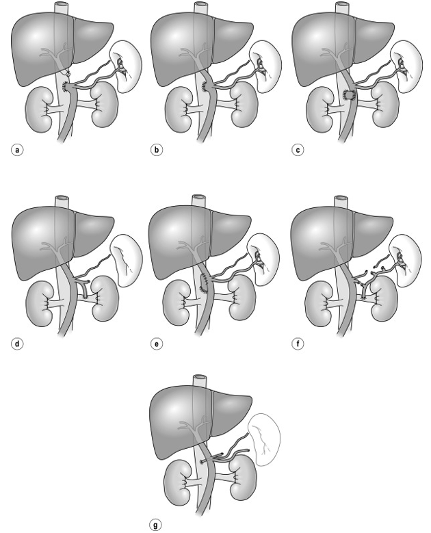 Figure
- non selective
- A - end-to-side portocaval
- B - side-to-side portacaval
- C - mesocaval
- D - proximal splenorenal
- Selective
- E - PTFE H-graft portocaval
- F - Distal splenorenal (Warren)
- Left gastric to IVC (Inokuchi)
Secondary prevention
- Main priority after initial haemorrhage controlled
- If no further therapy given, 70% re-bleed in 2 months
- Risk of re-bleed highest in hours-to-days after initial bleed
- Medical therapy to prevent rebleed
- Beta-Blocker (Nadolol)
- PPI to prevent ulcer
- Endoscopic management
- Repeat endoscopy and band ligation
- Every 10-14 days until varices eliminated
- Need patient compliance
- Consider
- Treatment of underlying liver disease
- Referral for transplant
- If recurrent bleeding despite endoscopic/medical management
- TIPS
- Liver transplant