Section: Hepatobiliary Sub-section: Pancreas Curriculum: Curriculum, page 89
Definition
Acute pancreatitis is a common disease, characterised by painful inflammation of the pancreas
Epidemiology
Incidence ~30/100,000
Aetiology
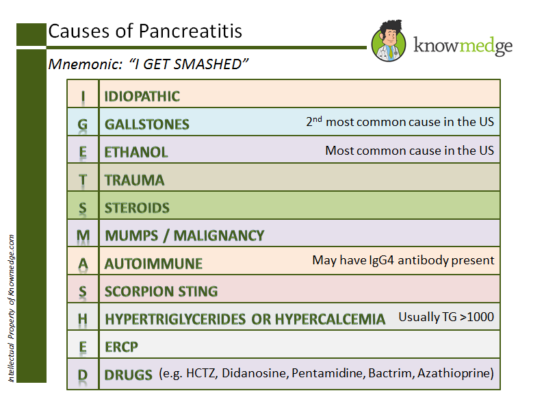
Gallstones
- EtOH
- Metabolic:
- Hyperlipidaemia
- Hypercalcaemia
- Iatrogenic: post ERCP, post operative
- Medication
- Steroids
- Other
- Trauma
- Autoimmune
- Genetic
- CF, PRSS1, SPINK1
- Anatomic
- Divisum
- Neoplastic
- Idiopathic
Pathology
- Normal
- Food
- Vagal nerves, vasoactive intestinal polypeptide (VIP), gastrin-releasing peptide (GRP), secretin, cholecystokinin (CCK), and encephalins stimulate the release of these proenzymes into the pancreatic duct.
- Proenzymes
- Travel to the brush border of the duodenum
- Trypsinogen hydrolysed by the brush border enzyme enterokinase to Trypsin
- Trypsin then converts other proenzymes into their active forms.
- Pancreatitis
- Injury to the acinar cells
- Intracellular activation of trypsin.
- Trypsin then acitives other proenzymes
- This triggers a local inflammatory r
- Release of both pro and anti-inflammatory mediators and chemokines.
- Results in microcirculatory disturbance including increased capillary permeability and microcirculatory intravascular coagulation and ischaemia.
- If inflammatory response is confined to a local response, this results in mild pancreatitis
- If systemic inflammatory response is mounted, then moderate or severe pancreatitis ensues. If sustained, MODS develops.
- After 7-10 days there is a transition through to a compensatory response syndrome with downregulation of immune system
- Explains why pancreatic infection peaks week 2 – 5.
Clinical
- Grey-Turner’s (flank ecchymosis) sign
- Often due to intraabdominal or retroperitoneal bleeding
- Pathophysiology
- In severe pancreatitis, injured pancreas release pancreatic enzymes that cause fat necrosis and inflammation and occasionally peri-pancreatic bleeding.
- This fluid can travel via an anatomical defect of the transversalis fascia to the space between the two layers of renal fascia. The bleeding then tracks to the pararenal area followed by the lateral edge of quadratus lumborum and finally to subcutaneous tissue of flanks.
- May not manifest for several days.
- Cullen’s (periumbilical ecchymosis) sign
- Often due to intraabdominal or retroperitoneal bleeding
- Pathophysiology:
- Haemorrhage tracks along gastrohepatic and falciform ligament to the umbilicus where it pools, leading to discolouration of abdominal wall and fatty tissue.
Diagnosis
- 2 of the folowing
- Imaging suggestive
- Lipase x3 upper limit of normal
- Severe upper abdominal pain
Initial management
- Fluid resuscitate and supportive care
- Co-existing cholangitis:
- SIRS + obstructive LFTs or dilated bile duct with gallstones on imaging can be diagnosed.
- IV abx and ERCP
- ERCP not indicated for acute pancreatitis without cholangitis
- Hypertriglyceridaemia (> 1000 mg/dL / > 55 mmol/L)
- Treatment with insulin infusion, anti-hyperlipidaemic medication, dietary manipulation and plasmapheresis
Severity assessment
SIRS
As per IAP/APA guidelines, use SIRS assessment:
| Variable | Criteria |
|---|---|
| Temperature | < 36’C or > 38’C |
| Heart rate | > 90 |
| Respiratory rate | > 20 |
| WCC | < 4 or > 12 |
| Transient SIRS < 48 hours | 8% mortality |
| Persistent SIRS > 48 hours | 25% mortality |
Revised Atlanta classification
- Mild - no organ dysfunction
- Moderatly-severe - transient (<48hours) of organ dysfunction, local or systemic complications
- Severe - persistent (>48hours) of organ dysfunction
CT imaging severity criteria (Balthazar criteria)
| CT grade | Grade score | Definition |
|---|---|---|
| A | 0 | Normal pancreas |
| B | 1 | Pancreatic enlargement |
| C | 2 | Pancreatic inflammation and/or peripancreatic fat |
| D | 3 | Single peripancreatic fluid collection |
| E | 4 | ≥ 2 fluid collections and/or retroperitoneal air |
| % of necrosis | Necrosis score | Definition |
| None | 0 | Uniform pancreatic enhancement |
| < 30% | 2 | Non-enhancement of region(s) of gland equivalent in size of pancreatic head |
| 30–50% | 4 | Non-enhancement of 30–50% of the gland |
| > 50% | 6 | Non-enhancement of over 50% of the gland |
| CT Severity Index | Morbidity | Mortality |
| 0–1 | 0 | 0 |
| 2–3 | 8% | 3% |
| 4–6 | 35% | 6% |
| 7–10 | 92% | 17% |
Ct severity index = grade + necrosis
Modified CTSI score
Easier to remember than CTSI:
| Pancreatic inflammation | 0: Normal 2: intrinsic abnormalities 4: fluid collection or peripancreatic fat necrosis |
|---|---|
| Pancreatic necrosis | 0: none 2: <30% 4: >30% |
| Extrapancreatic complications | 2: any of pleural effusion, ascites, vascular complication, parenchymal complication or GI involvement |
Total score:
- 0-2: mild
- 4-6: moderate
- 8-10: severe
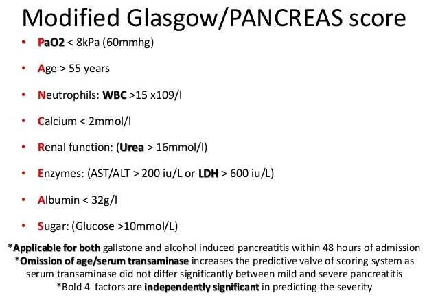
Glasgow-Imrie score
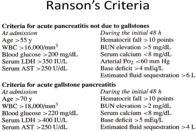
Ranson
Classification
Interstitial oedematous pancreatitis
- Majority
- Localised or diffuse enlargement of the pancreas is seen with homogeneous or slightly heterogeneous enhancement of the pancreatic parenchyma
- Complications include
- Acute peripancreatic fluid collection
- Pancreatic psudocyst
Necrotising pancreatitis
- 5-10%
- Three types defined by the Atlanta classification
- Pancreatic parenchymal necrosis alone,
- Peripancreatic necrosis alone
- Both (75-80%)
- Complications
- Acute necrotic collection
- Walled off necrotic collection
- These can be sterile or infected
Saxon Connor training day:
Early non-surgical interventions
- NSAIDS – may reduce MOF
- IV – moderate 5-10ml/kg/hr. Crystalloid solution
- Epidural analgesia – improves pancreatic perfusion, reduces need ventilation and mortality
- Intra-abdominal hypertension
- Neostigmine, perc drain.
- Decompressive laparotomy – high mortality, not indicated
- Nutrition – enteral where possible
- Early panc duct stenting – avoid
- Early lap chole – only in mild
- LMWH – 1mg/kg 12 hourly from admission 7 days, reduction in necrosis 26% to 6% - low level evidence.
- Insulin and plasmapheresis – PP has greater effect but costs more than insulin. Need RCTs.
- Thoracic duct lymph drainage – promising in Eastern Europe.
Indications for intervention in necroting panc
- Suspected or confirmed infected PN
- Persistent organ failure with walled-off necrosis
- Rare causes – ACS, bleed, gut ischaemia, gastric outlet or biliary obstruction due to mass effect
- Indications in sterile necrosis
Timing
- Should be delayed for 4 weeks
Intervention depends on:
- Timing
- Distribution of illness
- Systemic state of patient
MIRP (minimally invasive retroperitoneal pancreatic necrosectomy)
- Radiology guidewire along longitudinal axis of collection – if rigid scope, cannot access parts
- Operating theatre, dilate the tract under II
- Introduce nephroscope and remove necrotic tissue
- Continuous retroperitoneal lavage – be gentle, washout with saline, out through tract.
- Irrigating catheter (NG inside chest drain) left insitu
- Close monitoring by CRP
- Remove drains steadily on collapse of cavity.
Complications:
- Bleeding – primary (at time of procedure) or secondary.
- If blood in drain – could be sentinel bleed from pseudoaneurysm – needs urgent CT.
- Variceal – large foley catheter for tamponade, vasopressors, octreotide. PV stenting difficult, not really feasible.
- Biliary stricture, pseudocyst, pancreatic fistula
- Blocked drain
- Colonic necrosis – high mortality
- Biliary fistula – rare, due to erosion of intrapancreatic bile duct. May need PTC.
- Pancreatic fistula – MARP is to create a fistula. If proximal stricture and main duct intact then ERCP and stent.
- If disrupted main duct, may need pseudocyst drainage or resection of distal panc
- Upper GI fistula
- Erosion into duodenum. Optimise enteral feeding distal to fistula.
- Can result in recurrent collection
- Colonic fistula
- Local erosion by disease, thrombosis of mesocolic vessels, low perfusion state ischaemia
- Present with diarrhoea due to irrigation going into colon
- Treatment – irrigating drain with negative pressure, mostly close spontaneously. However high mortality.
- Biliary stricture
- Endoscopic with removable metal stents for 12 months, changed 3-6 months.
- Duodenal stricture
- Uncommon. May be associated with biliary stricture/fistula
- Delay repair until inflammation subsided. And perform gastrojejunostomy
- Pseudocyst
- Endoscopic 1st preference.
- Sometimes need to perc drain
- Portal vein thrombectomy/stent
- Delay by months. Let inflammation subside.
- For ascites that cannot be controlled.
- Anticoagulation