Section: Hepatobiliary Sub-section: Pancreas Curriculum: Curriculum, page 91
Epidemiology
5-10% post ERCP
Risk factors
- Patient-related: altered anatomy, female, older age, dilated CBD, presence of papillary lesion
- Procedure-related: sphincterotomy, use of precut needle knife, large balloon dilatation, stricture dilatation, difficult cannulation, long procedure, less experienced endoscopist.
Perforation
- 0.5-2%
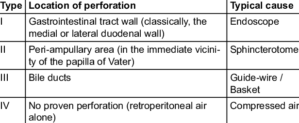
Grading:
- Mild – procedure aborted; LOS <4
- Moderate – LOS 4-10, ICU 1 night, RBC, intervention required
- Severe – LOS > 10, ICU > 1 night, surgery required, permanent disability
Management:
- Medical: NBM, IVF, IV Abx, NG decompression, possible TPN
- Endoscopic: stent (fully covered SEMS) or clip
- Usually type II managed with endoscopic biliary stent or PTC
- Surgery: type I usually need surgery
- Percutaneous drainage
Pancreatitis
- Most common
- May result from mechanical injury to the pancreatic duct, hydrostatic injury from contrast injection, or guidewire manipulation.
Prophylaxis
- Rectal NSAIDs (Diclofenac 100mg suppository)
- Pancreatic duct stenting
Bleeding
- 1-3%
- Optimize patients coagulation pre-op
- Usually can be managed with repeat endoscopy with injection and thermal coagulation or clip placement
- Fully covered metal stent placement is an option
Infection
- Incomplete drainage of an infected biliary system, obstruction of the cystic duct, infected pancreatic fluid collection, or rarely, contaminated endoscopic equipment