- Incision and exposure
- Make a longitudinal or gently curved incision over the ulnar aspect of the volar wrist, centered over the pisiform
- Incise skin and subcutaneous tissue
- Identify and protect the superficial branches of the ulnar nerve and cephalic vein (if encountered)
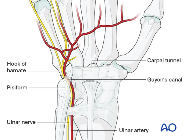
- Dissection and identification of Guyon’s canal
- Incise the palmar carpal ligament, the superficial roof of Guyon’s canal
- Identify the ulnar nerve and artery entering the canal lateral to the pisiform
- Carefully separate the ulnar nerve from the artery—they may share a fibrous sheath
- Decompression
- Divide the roof of the canal (palmar carpal ligament and, if needed, distal aponeurotic fascia of hypothenar muscles)
- Extend the release proximally and distally to fully decompress the nerve
- The nerve bifurcates in the canal:
- Superficial branch (sensory)
- Deep motor branch (dives deep through the hypothenar muscles)
- Release fascial bands overlying the deep motor branch as it travels deep to the hook of hamate and into the hand
- Be cautious to avoid injury to the ulnar artery, especially in trauma or aneurysmal cases
- Tendon transfers
- correct claw fingers
- possible grafts include ECRL, ECRB, palmaris longus
- tendons must pass volar to transverse metacarpal ligament in order to flex the proximal phalanx
- attach with either a two or four-tailed graft to the A2 pulley of the ring and small fingers
- restore power pinch
- Smith transfer using ECRB or FDS of ring finger
- restore adduction of small finger
- transfer ulnar insertion of EDM to A1 pulley or radial collateral ligament of the small finger
- transfer ulnar insertion of EDM to A1 pulley or radial collateral ligament of the small finger
- correct claw fingers
https://www.youtube.com/watch?v=qnH3lXa74LY&ab_channel=WUSTLLearnSurgery