Section: Head and neck Curriculum: Curriculum, page 43
Definition
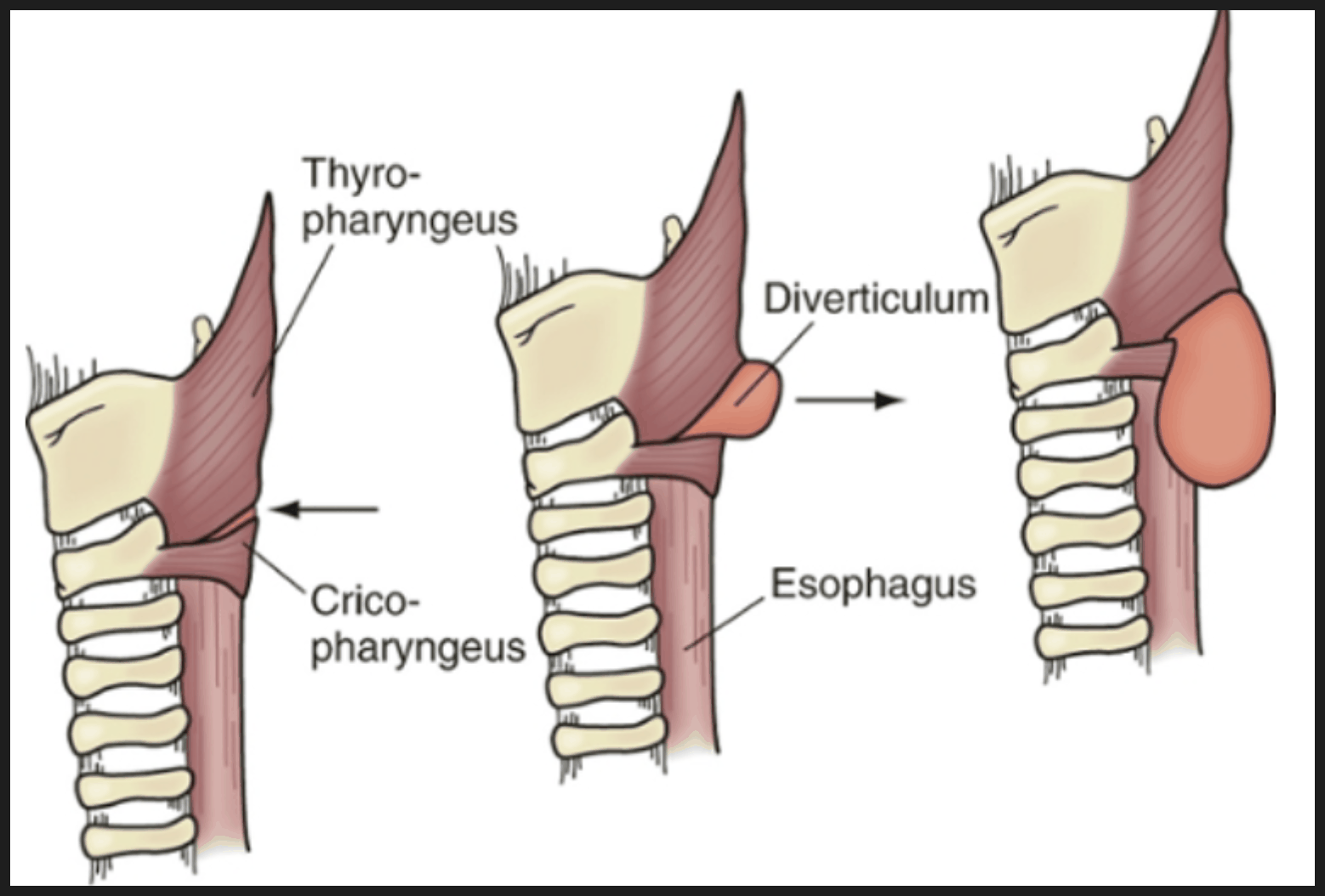
- A false pulsion diverticulum through Killians triangle
- Pulsion defect of the posterior mucosa between the two parts of inferior constrictor
- Killian’s triangle
- Oblique fibres of thyropharyngeus above
- Horizontal fibres of cricopharyngeus below
- Killian’s triangle
- With enlargement
- mucosal and submucosal layers dissect
- down left side of oesophagus
- into the superior mediastinum
- Posteriorly along prevertebral space
Incidence
- Most common type of oesophageal diverticulum
- M > F 3:1
- Most pts > 60yo; Rare < 30
Aetiology
- Loss of coordination during second stage of swallowing
- Simultaneous uncoordinated contraction of pharyngeal contractions and (pathologically) the cricopharyngeus sphincter
- “Herniation” through the unsupported triangular region devoid of muscle in the posterior wall
- With aging the sphincter muscle becomes non-compliant and fibrotic
- 1/3 of pts will have associated oesophageal functional / motility disorders
- Hiatus hernia, achalasia, diffuse oesophageal spasm
- Can also occur as part of a hereditary syndrome:
- Oculopharyngeal muscular dystrophy → Ptosis & Dysphagia
Histological
- Fibrosis, necrosis, atrophy, hypertrophy & inflammation
Clinical
- Dysphagia/ “Sticking in throat”
- Gurgling noises in the neck during swallowing
- Fetor oris / halitosis
- Regurgitation of undigested food; esp. when lying flat
- Respiratory complications – e.g. lung abscess, aspiration pneumonia
- Hoarse voice, pressure of the pouch on the RLN
- 30% of pts have associated GORD
- +/- may cause a swelling in the post triangle
- Below level of the thyroid cartilage
- Can be compressed / ‘emptied’
Investigations
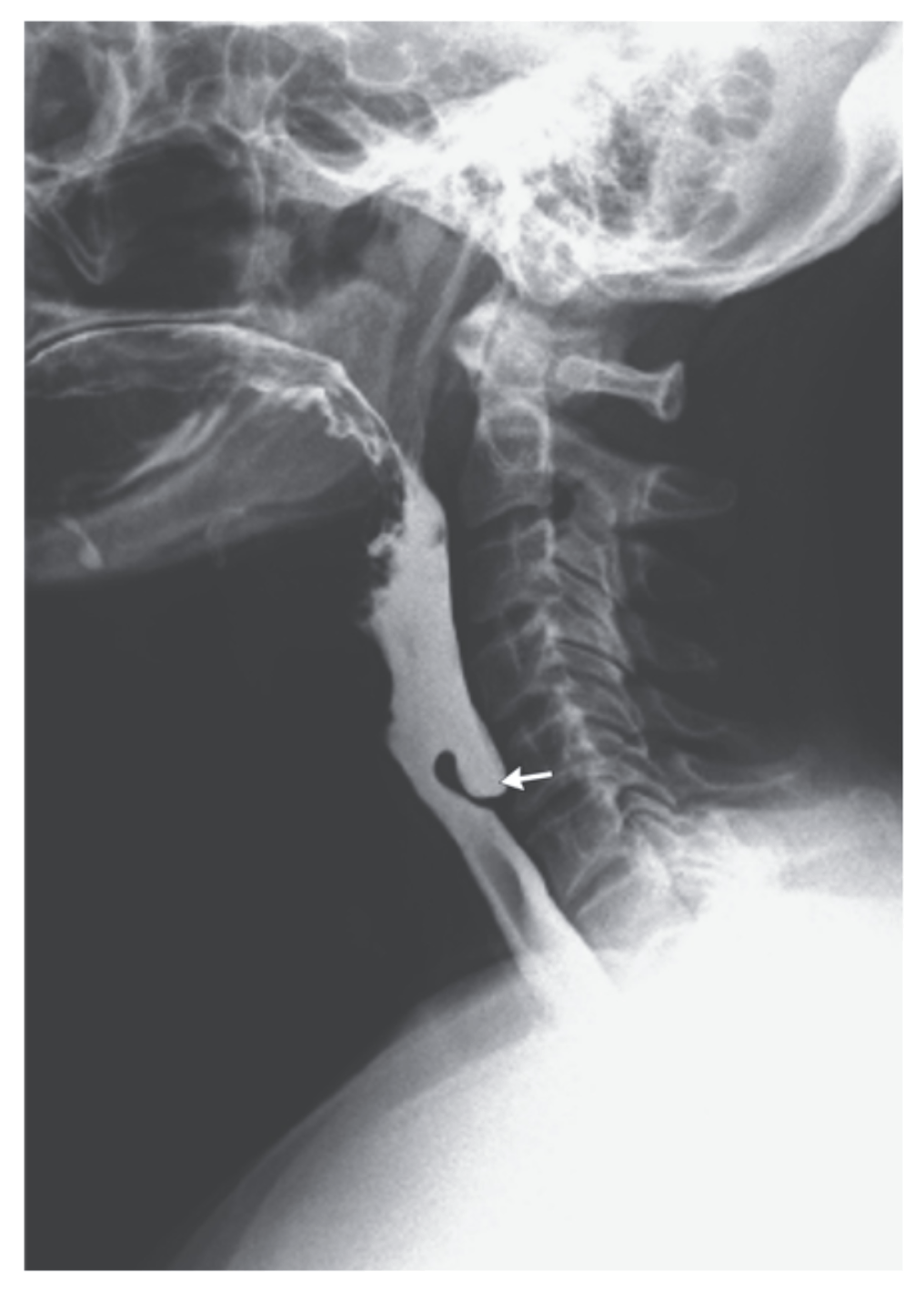
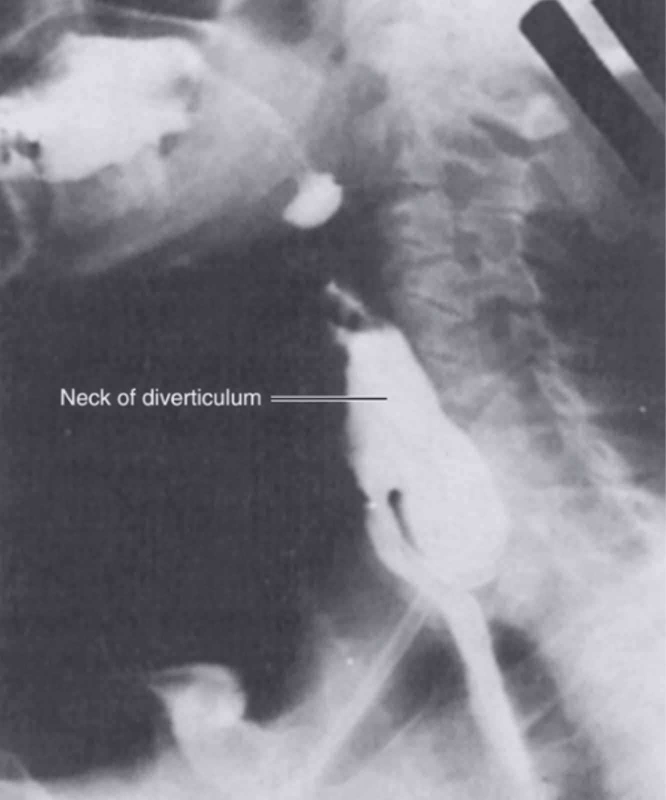
- Barium Swallow
- Diverticulum seen posteriorly at level of cricothyroid cartilage
- Lateral views critical in diagnosis
- Diverticulum seen posteriorly at level of cricothyroid cartilage
- Oesophageal Manometry (not required)
- Shows lack of coordination between pharynx and UOS
- +/- Hypertensive UOS
- Consider endoscopy if obstruction/malignancy suspected
- NB: Higher risk of perforation from scope in these patients
- CXR +/- Lung function tests
- Recurrent aspirations
Management
- Correct GORD first (before dividing the UOS) in order to avoid aspiration
Conservative
- Unfit for surgery
- Botox to cricopharyngeus
Endoscopic approach
- Dohlman’s procedure
- Oropharyngeally placed linear stapler/cutter to ablate the septum between the diverticulum & the cervical oesophagus
Surgery
- Cricopharyngeal Myotomy + Diverticulectomy or Diverticulopexy
- NB: Myotomy alone may be enough if < 2cm
- Get proximal and distal thyropharyngeus and cricopharyngeus
- Diverticulopexy = Suspend diverticulum upside down to posterior pharynx to prevent refilling
- Don’t suspend to prevertebral fascia as this will prevent movement with swallowing`
- NB: Myotomy alone may be enough if < 2cm
- For 3cm or smaller
- Open surgical repair superior to endoscopic
- For > 3cm
- Results equal for surgery and endoscopy
- Endoscopy has shorter hospital stay, and time to eating
- Results equal for surgery and endoscopy
Cricopharyngeal myotomy and diverticulectomy
- One stage cricopharyngeal myotomy and diverticulectomy has been the preferred approach in patients who are good surgical candidates
- Can be performed with a rigid endoscope or with an open cervical approach.
- Rigid endoscopes with specialised staplers can be used to divide the muscular bar and tissue between the Zenker’s and the oesophagus.
- NB: Need good neck extension (which some elderly don’t have!)
- In the open approach, the patient is positioned with the head and neck facing away from the affected side (most commonly left) and an incision is over the anterior border of SCM.
- The EJV may be ligated, the plane anterior to SCM developed with lateral retraction of the carotid sheath.
- The space between the carotid sheath laterally and the tracheo-oesophageal column medially is crossed by 3 structures;
- Omohyoid (transect or retract),
- Middle thyroid vein (ligate)
- Inferior thyroid artery (lower than the space needed)
- The diverticulum is then dissected free of surrounding tissue, opened, a finger inserted into the oesophagus, and the horizontal fibres of cricopharyngeus divided under vision.
- The diverticulum is then stapled off.
- The wound is closed in layers over a closed-suction drain.
Prognosis/ Natural Hx
- As diverticulum enlarges, tends to deviate from midline
- 90% to the left
- Likely due to the slight convexity of cervical oesophagus to left
- Also more laterally positioned carotid A on the left, creating a potential space for the sac
- 90% to the left
- Potential complications if untreated:
- Respiratory complications
- Ulceration
- Bleeding
- Formation of tracheo-oesophageal fistula
- Cancers occasionally occur in the pouch (SCC) ≈ 1%
- Results post-surgery (Mayo clinic data):
-
90% of pts asymptomatic
-
- Complications
- RLN palsy, cutaneous fistula, leak 5%
- Mortality 1.2%
Management of post-operative leak
- Investigations
- Gastrograffin (or Barium, if subtle) swallow
- CT neck and mediastinum
- With on table PO contrast
- Management
- IV ABs, IVF, NBM
- Drain collections (including in the mediastinum)
- Perc or open
- Place a feeding tube (fluoroscopically)
- Start feeding if stable
- Then wait for leak to seal (usually 3-4wks)