https://www.youtube.com/watch?v=4HIQF3RthBs&ab_channel=HoustonMethodistDeBakeyCVEducation
- Patient preparation
- Supine position
- General anaesthesia
- Prophylactic antibiotics (e.g. cefazolin unless contraindicated)
- Empty bladder with Foley catheter
- Port placement
- Open Hasson technique at the infraumbilical site
- Insert 10 mm camera port through a periumbilical incision
- Place a 5 mm working port in left lower quadrant (avoiding inferior epigastric vessels)
- Optional third port if adhesiolysis or omentopexy is required
- Inspection and assessment
- Evaluate peritoneal cavity for adhesions, hernias, or anatomical variations
- Perform adhesiolysis if needed for optimal catheter placement
- Introduce the catheter
- Skin incision just lateral or inferior to the umbilical port
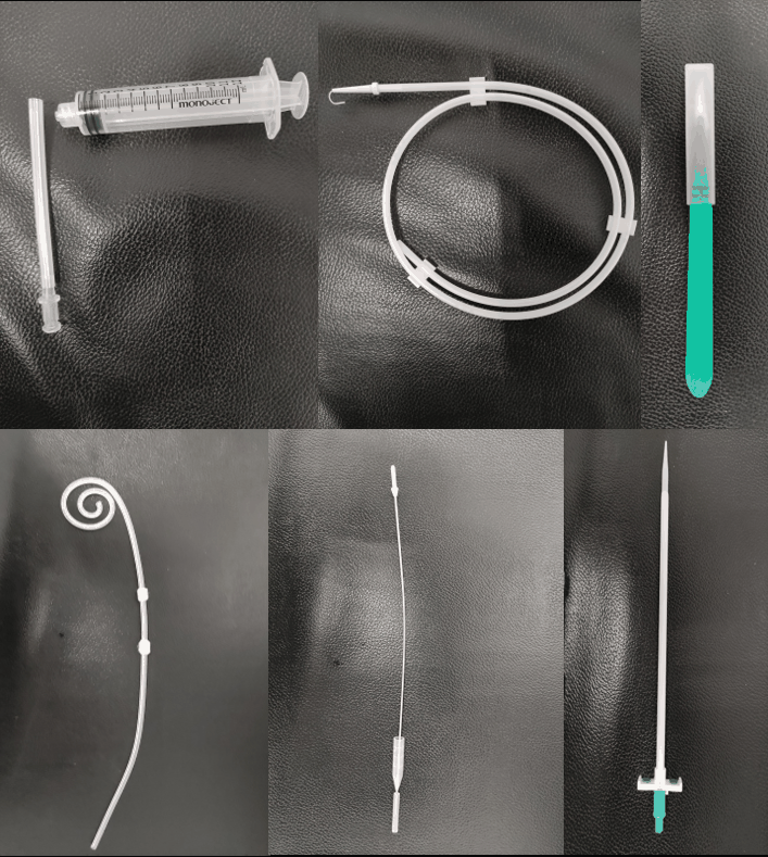
- Use a curled (Tenckhoff) PD catheter
- Insert the catheter into the abdomen through the deep entry point
- Advance it under laparoscopic vision to the pelvis
- Confirm that the curl lies freely in the pelvis, without kinking or looping
- Create the subcutaneous tunnel
- Select the exit site (typically in the left lower quadrant, lateral to rectus) — avoid belt line or skin folds
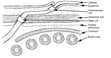
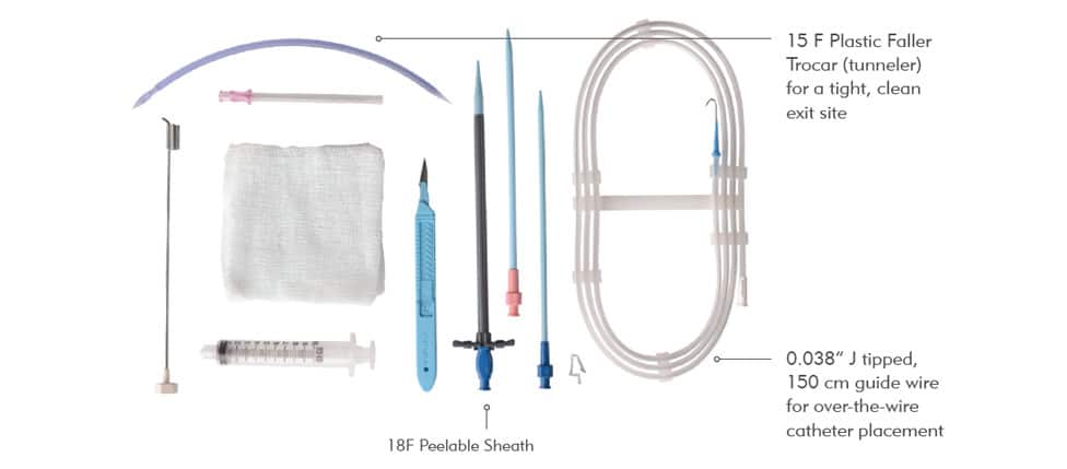
- Use a tunneller or long clamp to create a preperitoneal tunnel from the skin exit site to the deep cuff entry point (just lateral to the umbilicus)
- The tunnel should be gentle and curve inferior-laterally to reduce infection risk
- Secure and close
- Ensure deep cuff lies in the preperitoneal plane or rectus sheath
- Superficial cuff lies in the subcutaneous tissue near exit site
- Close port sites in layers
- Confirm catheter patency by flushing with heparinised saline
- Apply sterile dressing to exit site
- Post-operative care
- Delay use of catheter for 2–3 weeks to allow tract healing (unless urgent dialysis is required)
- Secure catheter and exit site to prevent tension
- Educate patient on PD technique and catheter care