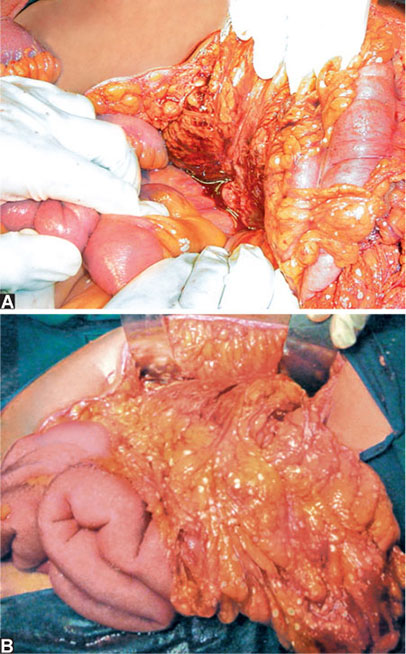
- Oedematous pancreas
- Appears swollen, boggy, and soft
- Loss of normal lobulated contour
- May have a dusky, pink-grey appearance due to interstitial oedema
- Peripancreatic fat stranding and necrosis
- Fat around the pancreas appears inflamed and streaky
- Fat necrosis (saponification)
- White, chalky deposits in mesentery or omentum
- Result of lipase-mediated fat breakdown and calcium deposition
- Serous or turbid fluid
- Free fluid in the lesser sac or around the pancreas
- May be straw-coloured, turbid, or blood-stained depending on severity
- Haemorrhagic areas
- Dark, bruised patches indicating vascular injury and necrosis
- Seen in severe or necrotising pancreatitis (may progress to pancreatic necrosis)
- Inflammatory adhesions
- Dense adhesions between pancreas and surrounding structures (e.g. stomach, colon, transverse mesocolon, omentum)
- Dissection may be difficult and bloody
- Pancreatic necrosis (in severe cases)
- Patchy or confluent areas of black or green-grey devitalised pancreatic tissue
- May be sterile or infected
Saponification