Section: Vascular Curriculum: Curriculum, page 66, Curriculum, page 70
Key principles
- Typically for long-term venous access e.g. Chemotherapy or dialysis
- Typically use subclavian vein, however can also use IJV
- Hickmann and Port-a-cath insertion follow similar steps, however, a port-a-cath has a port placed in a subcutaneous pocket which can be accessed via a Huber needle
Operation Details
- Typically performed under GA. IV Cephazolin
- Prep and drape
- Flush all device used with heparinised saline heparinised saline (10 units/mL) - place 5000U into a 500ml bag of NaCl
Subclavian
- Patient supine with a roll beneath shoulder blades.
- Head up neck turned to contralateral side
- Identify landmark- junction of outer 1/3 and middle 1/3 clavicle
- Cannulation of the SCV using a Seldinger tecnhque, needle towards opposite shoulder, walk down under clavicle, bevel downwards and advance until flashback
- Place guidewire placed watching for ventricular ectopics under II guidance
- Remove needle
- Small skin incision
- Advance dilator over wire
- Remove dilator
- Advance catheter over guidewire
- Usally 15cm on right and 20cm on left
- Check position with XR
- Secure catheter and place sterile dressing
Internal jugular vein
- Anatomical landmarks
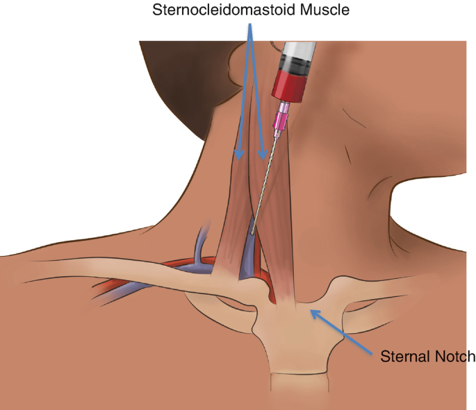
- Internal jugular vein lies lateral to carotid artery, within the carotid sheath
- Right IJV preferred (straighter path to SVC, lower risk of thoracic duct injury)
- Common landmark: apex of triangle formed by the sternal and clavicular heads of sternocleidomastoid (SCM), just above the clavicle
- Needle directed caudally, slightly laterally, and posteriorly toward ipsilateral nipple
- Steps
- Patient supine in 15–20° Trendelenburg (reduces air embolism, distends vein)
- Turn head slightly to the contralateral side
- Sterile prep and drape; local anaesthetic infiltration at puncture site
- Use either landmark technique or ultrasound guidance (preferred)
- Insert introducer needle at apex of triangle, lateral to carotid pulsation
- Advance with aspiration until dark venous blood is aspirated
- Insert guidewire through needle
- Remove needle, make small skin incision
- Advance dilator over guidewire, dilate soft tissue
- Remove dilator, pass catheter over wire to appropriate depth (usually ~15 cm)
- Remove guidewire, aspirate and flush catheter ports
- Secure catheter with sutures, apply sterile dressing
- Confirm position and rule out complications with chest X-ray
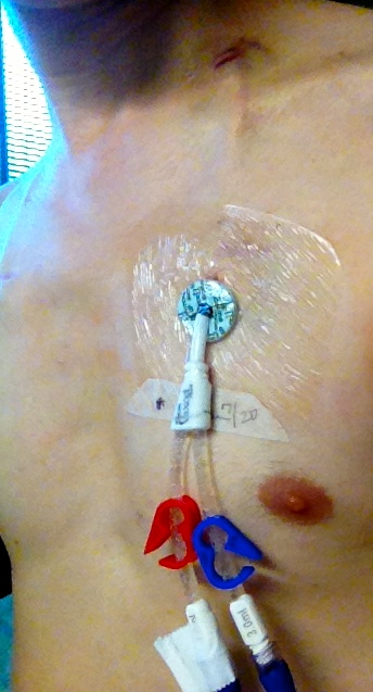
Hickman line insertion
- Vein cannulation
- As above - either IJV or SV, typically IJV
- Canulation → guidewire → dilate
- Insert peel-away sheath over guidewire
- Exit site
- Usually 5–10 cm below clavicle)
- Create a subcutaneous tunnel from the exit site to the cannulation site
- Use tunnelling device to pull catheter through the tunnel and out the first cannulation site
- Ensure cuff lies in the tunnel ~2 cm from exit site
- Dacron cuff to allow tissue ingrowth
- Advance catheter through peel-away sheath into SVC (tip at cavoatrial junction)
- Remove sheath, confirm catheter flow and flush all lumens
- Secure at skin with sutures or securement device
- Apply sterile dressing
- Chest X-ray to confirm tip position and exclude pneumothorax
https://www.youtube.com/watch?v=PvQXthhhl7I&ab_channel=VascularAccess
Port-a-Cath insertion
- This is the same process as above with a similar second incision 5-10cm inferior to the clavicle
- Steps
- Venous access
- As above
- Pocket is created
- Pocket is created on the chest wall, exposing the pectoral fascia.
- Subcutaneous tunnel created
- Catheter passed to venous access site and placed via the peel-away sheeth
- Port fixation
- The infusion port is fixed to chest wall by placing sutures that pass through the flanges of the port onto the fascia of the chest wall in 4 places to ensure that the port will not flip.
- Venous access
- The skin is closed
- Port accessed and locked with heparinized saline
Intraoperative complications & challenges
• Arterial cannulation or unable to cannulate SCV o Try contralateral side o Try IJV o Convert to cephalic vein cut down to place port
Post-operative complications
• Immediate o Pneumothorax – 1-3% - may need chest drain o Air embolism – administer high flow O2 o Bleeding o Arrhythmia o Rarely pericardial tamponade • Early o Infection o AV fistula • Late o Catheter migration o Thrombosis o Line sepsis o Fracture o Failure
Specific post-operative care
• CXR in recovery • Access as required
https://www.youtube.com/watch?v=-jWMYh0Y6gg&t=17s&ab_channel=IREducation