Section: Trauma Curriculum: Curriculum, page 77
Classification
TBI Severity
| Mild | Moderate | Severe | |
|---|---|---|---|
| LOC | Brief (sec - min) | < 24hrs | > 24hrs |
| PTA (Amnesia) | < 1hr | 1 - 24hrs | > 24hrs |
| Imaging | Normal | Abnormal | Abnormal |
| GCS | 13-15 | 9-12 | 3-8 |
- Severe Traumatic Brain Injury
- LOC and post traumatic amnesia > 24hrs with abnormal imaging and GCS 8 or less (tubed)
- Pathological Classification
- Focal
- Diffuse → DAI
- Blunt vs penetrating
Pathophysiology Raised ICP
- Measuring Physiological Parameters in TBI
- MAP - SBP + 2xDBP/3
- ICP
- Normal: 7-15mmHg
-
20mmHg - Mass lesion (haematoma/oedema) → Decompress
- Affected by - CSF Volume, brain water, CBV, Cerebral VR
- Broadly affected inflow, outflow, cerebral oedema, CSF fluid volume
- Cerebral perfusion pressure (CPP)
- Net pressure gradient driving cerebral blood supply
- MAP-ICP = CPP - normal is 60-70mmHg (supine)
- Cerebral blood flow (CBF)
- Normal - 50-55ml/min/100g brain tissue (15% CO)
- Tightly autoregulated
- BP, PaO2, PaCO2
- Pathophysiology of TBI
- Direct tissue damage and impaired CBF regulation
- Lactic acid build up via anaerobic respiration
- Cerebral oedema via increase membrane permeability
- Cerebral Perfusion Pressure (CPP) = MAP - ICP
- Increased ICP causes reduced CPP
- Leads to cerebral ischaemia and brain injury
- CPP of 60-70mmHg recommended after severe traumatic brain trauma
- Cerebral autoregulation controls CPP normally but after trauma it can function abnormally
- When autoregulation mechanism is intact
- Raised ICP causes lower CPP
- Vasodilation occurs to increase CBF and CPP (increased compliance)
- When autoregulation mechanism is intact
Management of TBI
- In TBI, the normal tight autoregulation of CBF is compromised
- Principles of Management
- Maintenance of BP and oxygenation
- Administer oxygen and usual resuscitation with target MAP 90-100
- Reducing secondary hits
- Treat and prevent
- Anaemia
- Hypo/hyperglycaemia
- Hyponatremia
- Fever
- Treat and prevent
- Adjuncts
- ICP monitoring
- ICP treatment
- Mannitol - causes hypovolaemia
- Hypertonic saline
- Barbiturates if refractory
- Hyperventilation
- Maintenance of BP and oxygenation
Surgery - Burr hole and Craniectomy
- Indications for rural neurosurgical intervention
- Neurologically deteriorating pt and can’t do CT head
- T/F to nearest N/S unit is > 2 hours away
- Sizable intracranial haematoma
- Indications for Burr hole
- GCS 8 or less with imaging showing epidural haematoma causing midline shift and unequal pupils
- High clinical suspicion with palpable #, ipsilateral fixed dilated pupil and deteriorating neurology without imaging
- Burr Hole technique
- Confirm side and position → Shave and prep
- LA and 3cm incision to bone
- Periosteal elevation and self-retainer
- Hudson brace manual burr used perpendicular to skull with saline being applied for bone dust
- Stop when resistance gives
- Perforator for conical opening widened by cylinder burr
- Epidural clot should evacuate now
- If it doesn’t = Subdural → Open dura with knife
- If no blood - confirm side/scan and if bleeding on-going DO NOT TAMPONADE
- Transfer patient
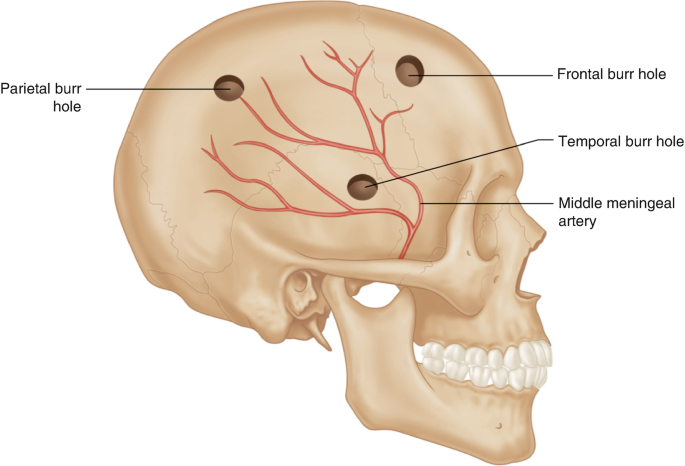
- Locations:
- Temporal - 1cm above Posterior Zygomatic Arch
- Frontal - 10cm above pupil - over the Coronal Suture approx 10cm cephalad and in line with the Mid-Pupillary line
- Parietal - 5cm above ear - over the Parietal Eminence
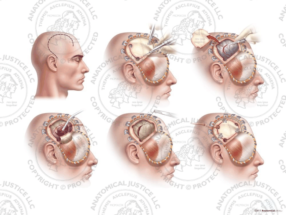
- Emergency Frontotemporal Craniectomy
- Scalp incision from ant to ear curving up and anteriorly to follow temporal plane
- Raise flap
- Serial burr holes include edge of # and combine with Gigli saw
- Elevate bone flap
- Don’t dig out bone fragments from bone parenchyma - causes more injury
- Death occurs due to on-going bleeding → High ICP → Brain shift and herniation causing brain stem damage