Section: UGI Sub-section: Gastric Curriculum: Curriculum, page 96
- Arise from Enterochromaffin-like Cells
- Located in Gastric Fundus or Body
- Neural crest origin
- Base of Crypts of Lieberkühn
- Rare
- <1% of gastric neoplasms, 8% of all GIT NETs
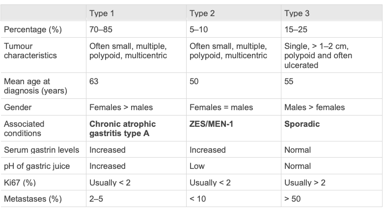
Type 1 Gastric NETS
- 70-80% of Gastric NETs
- Older pts, F > M 3:1
- Associated Chronic Atrophic Gastritis
- Achlorhydria and Vit B12 malabsorption (Pernicious Anaemia)
- Decreased acid, pH, leads to increased gastrin
- Causes hyperplasia of ECL cells
- NB: CAG very common in elderly, only 1% develop NETs
- Achlorhydria and Vit B12 malabsorption (Pernicious Anaemia)
- In fundus/body, frequently multicentric
- Mostly small, Rarely > 2cm
- Small lesions almost always benign
- 1cm predominantly benign, <10% invasion
- If invasive, behave well, low risk mets (5% to LNs, 2% distant)
- Very rare to die from this
Type 2 Gastric NETS
- 6-8% of gastric NETS
- Associated with MEN1 and Gastrinoma causing ZES
- F=M, age 45-50
- NB: ECL hyperplasia in 80% of MEN1 ZES
- 5-30% of these develop gastric NETs
- Hyperplasia, dysplasia, neoplasia sequence
- Hypergastrinaemia and MEN1 gene abnormality seemingly needed for gastric NET formation
- Very rare to have gastric NETs in ZES that are sporadic and not caused by ZES
- Very rare to have gastric NETs in MEN1 patients that don’t have ZES
- Located body and fundus
- Mostly multiple and small, occasionally larger
- Malignant potential intermediate
- 90% non-invasive
- But if invasive, 30% LN mets, 10-20% distant mets
- NB: Potential for other tumours related to MEN 1 to have caused the metastases
- Overall prognosis usually depends on other MEN 1 related tumours
- Prognosis for type 2 NETs is favourable
Type 3 Gastric NETS
- 15-20% of gastric NETs
- Sporadic, no association to hypergastrinaemia
- Non-atrophic gastric mucosa
- M > F 3:1, Age 50yrs
- Usually solitary
- Often large 70% > 1cm, mean 3.2cm
- Much more aggressive than Type 1 and 2
- Many already disseminated by diagnosis
- LN metastases in 71%
- Liver metastases 69%
- Total survival 50% at 5yrs
- If distant metastases – 10% 5yr survival
- Mostly well differentiated, often G2 (Ki67 > 2%)
- Atypical carcinoid syndrome in 5-10%
- The flushes may be patchy, sharply demarcated, serpiginous, and cherry red; they are also intensely pruritic. Diarrhea or cardiac lesions are unusual. The tumors that cause this variant syndrome secrete histamine
- Tumour histamine release – breakdown product MelmAA in urine as tumour marker
- Itching, cutaneous oedema, bronchospasm, salivary gland swelling, lacrimation
- Very few have serotonin release
- Urinary 5-HIAA not appropriate tumour marker
Diagnosis
- Most found incidentally
- On scope for anaemia or abdo symptoms
- Can bleed or have gastric outlet obstruction if large
- Hx
- ? Chronic atrophic gastritis,
- ? MEN1 related malignancies (3Ps)
- Bloods
- Gastrin, MEN1 screen
- Gastroscopy
- Biopsy of lesion and 2 x antral, fundus, body
- Look for ECL Hyperplasia, Atrophic Gastritis
- EUS for
- Types 1 & 2 > 1cm
- All type 3
- Staging – CT CAP
- Octreotide scan can be considered in well differentiated tumours
Management
- Type 1 – Chronic Atrophic Gastritis
- Follow yearly if < 1cm
-
1 cm with no invasion – EMR
- If larger or invasive, surgical excision
- Malignancy – partial/total gastrectomy with lymphadenectomy
- Type 2 – Gastrinoma/Zollinger Ellison
- Need to remove source of gastrin (gastrinoma) and the NET
- Open exposure, both duo and pancreas seen via duodenotomy - 80% distal pancreatectomy most often performed
- Local excision of the gastric NET also unless large
- Larger get Gastric resection and lymphadenectomy
- Type 3 Sporadic
- Risk of malignancy even if small
- Most require gastric resection with lymphadenectomy
-
2cm consider gastrectomy
- If mets, tumour debulking may alleviate symptoms
- Hepatic mets – resection/Hepatic A embolisation/TACE/RFA
- Octreotide for symptomatic palliation
2023 ENET Management guidelines
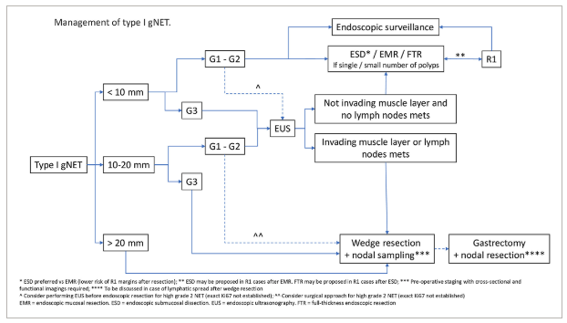 Type 1
- <1cm observe
- 1-2cm gray zone
-
2cm resect
Type 2 “Type II gNETs are the rarest type of gNET (5% of cases). They arise in the context of hypergastrinaemia associated with ZES which may occur in patients with MEN-I. Treatment of patients with type II gNETs strictly depends on the management of the MEN-I syndrome.”
Type 3
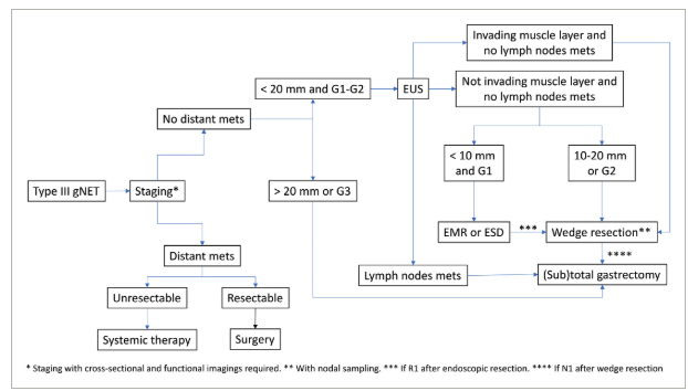
- All require resection
- Option based on size, grade and depth of invasion