Section: Urology Curriculum: Curriculum, page 6
Incidence
- 3.7/100,000
- More common NZ, less common Africa/Asia
- Peak incidence 15-35
- Teratoma 20-30
- Seminoma 30-40
- Yolk sac younger (90% < 15)
- Bilateral tumours 5%
Classification
- Germ cell 85%
- Seminoma
- Non-Seminoma
- Teratoma (has cells from more than one germ cell layer)
- Choriocarcinoma -Syncytiotrophoblasts and Cytotrophoblasts
- Embryonal
- Yolk Sac
- Sex cord-stromal tumours
- Sertoli cell tumour
- Leydig cell tumour
- Granulosa cell tumour
- Mixed types e.g. sertoli-leydig cell tumour
- Unclassified
- Mixed germ cell and stromal tumours
- Gonadoblastoma
- Adnexal and paratesticular tumours
- Adenocarcinoma of the rete testis
- Adenocarcinoma of the epididymis
- Mesothelial neoplasms
- Malignant mesothelioma
- Adenomatoid tumour
- Miscellaneous tumours
- Carcinoid
- Lymphoma
- Metastatic tumours (prostate carcinoma is the most common)>)
Risk Factors
- Cryptorchidism – orchidopexy does not stop risk
- Genetic risk
- Testicular dysgenesis, Kleinfelter’s, testicular feminization
- Contralateral tumour – 5% cumulative risk
Clinical
- Painless testicular mass, not separable from testis
- 40% present with tenderness which may reflect small secondary hydrocele
- Gynaecomastia
- 30% present with metastatic disease
Pathology
- Seminomas – Lymphatic spread to Retroperitoneal lymph nodes
- Choriocarcinoma – haematogenous spread
| Feature | Seminoma | NSGCTs |
|---|---|---|
| Pathology | Uniform germ cells with clear cytoplasm, lymphocytic infiltrate. | Includes embryonal carcinoma, teratoma, choriocarcinoma, and yolk sac tumors, showing diverse differentiation. |
| Age Group | Common in men aged 30–40 years. | Common in younger men aged 15–35 years. |
| Growth and Spread | Remains localised. Slow-growing; late lymphatic spread. | Faster-growing; early metastasis to lymph nodes and distant organs. |
| Lymph Node Spread | Retroperitoneal lymph nodes. | Retroperitoneal and distant lymphatic spread. |
| Hematogenous Spread | Rare, occurs late. | Common, especially in choriocarcinoma (lungs, liver, brain). |
| Tumor Markers | β-hCG (mildly elevated in ~10–15%); no AFP. | 70% = May have elevated AFP (yolk sac or mixed tumors), β-hCG (choriocarcinoma or mixed tumors). |
| Management | - Stage I: Orchiectomy, surveillance, or carboplatin. - Stage II-III: Radiation or BEP chemotherapy. | - Stage I: Orchiectomy, surveillance, or BEP chemotherapy. - Stage II-III: BEP chemotherapy. |
| Radiation Sensitivity | Highly radiosensitive. | Poorly radiosensitive; managed primarily with chemotherapy. |
| Prognosis | Excellent; 5-year survival >95% in early stages. | Variable; depends on type and stage, but generally more aggressive than seminomas. |
| Relapse Risk | Lower, particularly in early stages. | Higher risk of recurrence, especially with mixed components. |
Investigation
- Blood tests – aFP (yolk sac cells), hCG (trophoblasts), LDH
- USS to investigate testicle
- Seminoma - well-defined hypoechoic lesions without cystic areas
- Non-seminomatous germ cell tumours - non-homogenous hyperechoic lesions with calcs, cystic areas and indistinct margins
- CT abdo/pelvis + CXR
- DO NOT FNA (risk of disrupting lymphatics)
Staging
T1: Tumour limited to testis & epididymis, without LVI
T2: Tumour as above but with LVI or extending through to tunica vaginalis
T3: Tumour invades spermatic cord
T4: Tumours invades the scrotum
N1: LN mass < 2cm N2: LN mass 2-5cm N3: LN mass >5cm
M1 - distant nets
S0 - Markers within normal limits S1 - HCG <5,000, αFP <1,000, LDH <1.5x S2- HCG 5,000-50,000, aFP 1,000-10,000, LDH 1.5-10x S3- HCG >50,000, aFP >10,000, LDH >10x
M1a non-regional nodal or pulmonary M1b distant
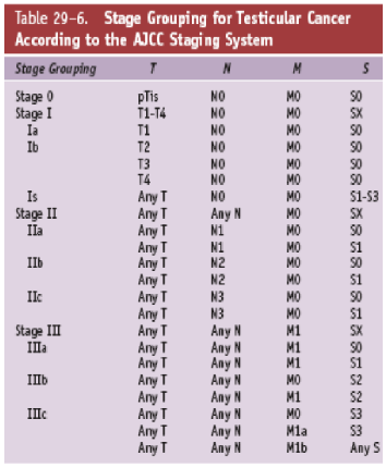
Summary of staging
- Stage 1 = no LN
- Stage 2 = LN positive
- Stage 3 = distant mets
Management
| Feature | Seminoma | NSGCTs |
|---|---|---|
| Stage 1 | Orϲhiеϲtоmy + Surveillance | Orchiectomy alone in low risk disease (no LVI, not embryonal carcinoma, >T2) In high risk patients consider BEP or RPLND |
| Stage 2 | + RTx or Chemo | Clinical + RPLND or BEP – BEP preferred in IIB/C Pathological BEP |
| Stage 3 | Chemo | Chemo |
Principles of management
- Resection of primary for local control, confirmation of histological diagnosis and prognostication
- Then treatment based on stage
- Seminoma
- Most will receive radiotherapy
- Consider chemotherapy
- Non-seminoma
- Most will receive RPLND
- Consider chemotherapy
- Seminoma
Inguinal orchidectomy
- Diagnosis and prognostication
- How to
- 2cm skin incision superior and parallel to inguinal ligament
- Through EO in line of fibres
- ID and preserve ilioinguinal n
- Dissect spermatic cord free along its length and isolate it at PT
- Stretch cord opening and deliver the testicle
- Divide the spermatic cord in 2 parts - vascular and vas, close to deep ring
- Layered closure
Seminoma
- Radiotherapy
- Seminomas are more radiosensitive
- Usually given to retroperitoneum
- 30Gy to para-aortic and ipsilateral iliac LN
- Chemotherapy Options
NSGCT
- Prophylactic retroperitoneal LN dissection
- Chemo
- cisplatin/bleo regimes - 85% cure even with mets
- Close F/U
Steps in management and MDM points
- Confirmation of diagnosis - usually with radical orchidectomy
- Risk stratification
- Histo - seminoma vs non-seminoma
- Mets or not
- Serum tumour marker degree of elevation
- Semen cryopreservation
- Baseline sperm count and banking pre dx and staging
- Infertility risk from all mx options (surgery, RTX) and the pathology
- Specific management plans then differ based on the stage of disease and histology
- This can include
- Retroperitoneal lymphadenectomy
- Chemotherapy
- Radiotherapy
Follow-up
- Self examination 3-6 monthly
- Annual examination
- Serum hCG and AFP useful for NSGCT but nothing required for seminoma
Natural history
- Testicular GCTs are very curable with 95% 5 yr survival and often present with limited local disease (particularly seminoma)
- When LN mets do occur it usually goes to para-aortic LNs so imaging needs to assess the retroperitoneal LNs
- Mixed cancers get treated as NSGCT
Prognosis
- LN neg disease = 100% 5 yr survival
- Poor prognostic features
- Markers
- AFP >10000, hCG >50,000, LDH >10 (10x upper limit of normal)
- Markers