Section: Abdominal wall and retroperitoneum Sub-section: Hernias Curriculum: Curriculum, page 2
Definition
- A hernia that passes into the Femoral canal
Incidence
- 7% of abdominal wall hernias are femoral
- But in both sexes, femoral hernia is less common than inguinal (1:25)
- More common in female
- Elderly, associated with weight loss/changes in fat distribution
- Esp. women who have borne children
- Account for ≈ 1/3 of groin hernias in women
- 2% in men
- More common on RHS (2:1)
- Bilateral in up to 20%?
Classification
- Classical – through femoral canal
- Pre-vascular
- Under inguinal ligament, in front of femoral artery and vein
- Wide, flattened neck
- Difficult to repair
- May need mesh plug
Aetiology
- Predisposing factors:
- Female pelvic anatomy
- Obesity
- Pregnancy
- Increased intra-abdo pressure
- Chronic cough, straining / constipation
Clinical
- Mostly asymptomatic until incarcerated/strangulated
- Present as an irreducible swelling at the femoral triangle
- Can be indistinguishable from a node or a synovial cyst
- Usually appear irreducible, even with an empty sac, because of associated fat and nodes
- Rarely they can be fully eliminated with manipulation, in which case they can be confused with a saphenous varix (dilation of great saphenous vein at junction with femoral vein)
- Richter’s type femoral hernias
- May present late with minimal local signs and bowel obstruction/infarction
- De Garengeot Hernia – Appendix in femoral hernia sac
Pathology
- Preperitoneal fat is forced out of the canal, followed by adherent peritoneum
- Femoral ring wider in women, as the iliopubic tract inserts into Cooper’s ligament
- M+M due to toxaemia that results from strangulation
Management
- Operative Technique
- Three classical approaches to the femoral canal have been described
- Low (Lockwood) - Elective
- Transinguinal (Lotheissen)
- High (McEvedy) – Incarcerated/strangulated
- Three classical approaches to the femoral canal have been described
- Key operative steps:
- Dissection of the sac
- Reduction / inspection of the contents
- Ligation of the sac
- Approximation of the inguinal and pectineal ligaments
Modified McEvedy - High Approach
- Incision
- Transverse skin crease incision 3cm above pubic tubercle
- 8 cm long to midline
- Ligate Superficial Epigastric Veins
- Entry
- Divide the rectus sheath a few cm from its lateral boarder
- Sweep rectus medially
- Divide the transversalis fascia
- Identify inferior epigastrics, ligate if necessary
- Dissect down on peritoneum to femoral ring
- Management of the hernia
- If incarcerated, I would routinely open the peritoneum to control spillage with the use of soft bowel clamps before reducing
- Reduce hernia with pressure from below
- If doesn’t reduce easily
- Follow external oblique inferiorly, dissect out inguinal ligament
- Dissect out sac, open & inspect contents
- Reduce or incise neck
- If need to incise ring then relaxing incision in inguinal ligament
- Lacunar ligament incision runs the risk of bleeding from Aberrant Obturator Artery
- Ensure contents inspected
- Bowel resection if necessary
- Repair
- Close femoral canal with 2-3 ethibond /PDS sutures
- Finger over femoral vein to protect (not retracting just overlying so as not to narrow vein)
- Suture iliopubic tract to pectineal ligament
- Closure
- Close Anterior rectus sheath with 0 maxon. S/c 3-0 monocryl to skin
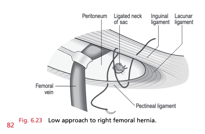
Lockwood - Low Approach
- Incision
- Groin crease incision below the medial half of the inguinal ligament
- 5 cm long over lump
- Dissection
- Dissect onto hernia
- Define neck of hernia under inguinal ligament
- Hernia
- Open sac, inspect contents
- Reduce contents
- If necessary, enlarge ring, incise neck, or relaxing incision in inguinal ligament
- Define neck
- Transfix neck of sac & excise sac
- Repair
- Close femoral canal with 2-3 Nylon or Ethibond sutures
- Interuppted
- Check the vein is not occluded before tying
- Finger over Femoral Vein to protect
- Suture inguinal ligament to the pectineal ligament
- Consider mesh plug as an alternative
- Close femoral canal with 2-3 Nylon or Ethibond sutures
- Closure
- S/C 3-0 monocryl to skin
- S/C 3-0 monocryl to skin
If the femoral vein is torn, control the bleeding with pressure from gauze packs for 5 minutes. Meanwhile, order blood, arterial sutures, tapes, bulldog clamps and heparin solution, and summon assistance. Expose the vein; do not hesitate to approach it from above and below the inguinal ligament. Apply bulldog clamps and tapes above and below the damaged segment. Insert fine 5/0 sutures set 1 mm apart, 1 mm from the torn edges, to evert them and close the hole. Flush with heparin at intervals. Release, then remove the clamps and tapes
Lothiesen - transinguinal approach
- Advantage of both inguinal and femoral
- Entry
- As per inguinal hernia
- Incside transveralis fascia
- Hernia
- Identify sac and external iliac vein
- Gently reduce
- Transfix and ligate
- Repair
- Close the femoral canal with 2-0 non- absorbable suture
- Closure
- Either close the transversalis fascia with non-absorbable suture or perform a mesh repair