Overview
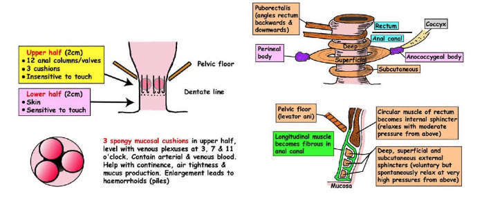
- Last 4cm of the alimentary tract
- Usually shorter in females
- Tube of muscle but all fibres are circular consisting of the Internal and External Anal Sphincters
- Internal Anal Sphincter – Visceral muscle
- External Anal Sphincter – Skeletal muscle
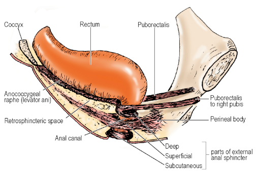
- Junction of Rectum and Anal Canal:
- At the Pelvic Floor
- At the level where the Puborectalis part of the Levator Ani clasps the gut and angles it forwards
-
Located 2.5cm anterior to the tip of the Coccyx
-
Muscles of Anal Canal
- ‘A tube within a funnel’
- Sides of the upper part of the funnel – Levator Ani muscles
- Stem of the funnel – External Anal Sphincter continuous with the Levator Ani muscle
- Tube within the funnel – Internal Anal Sphincter – thickened continuation of the inner circular layer of the rectal muscle
- Internally lies the Submucosa and Mucous membrane
-
Corrugator Cutis Ani muscle – separate smooth muscle fibres in the peri-anal skin causing puckering of this area
External Anal Sphincter
- Deep, superficial and subcutaneous parts
- Upper (Rectal) end
- Circular skeletal muscle fibres blend with the Puborectalis part of the Levator Ani except in the midline at the front (where there are no Levator Ani fibres)
- Region where Puborectalis fuses with the External Sphincter is termed the Anorectal Ring
- Also the level of the upper end of the Internal Sphincter
- Palpable on rectal exam
- Middle part
- Fibromuscular strands pass backwards to the posterior surface of the Coccyx contributing to the Anococcygeal Ligament
- Retrosphincteric space occupied by fibrofatty tissue lies between these fibres and the muscular Raphe formed by the Iliococcygeal part of the Levator Ani
- Post-Anal Plate = Multi-layered fibromuscular Anococcygeal Ligament (with External Sphincter, Iliococcygeus and Pubococcygeus components) + Overlying Superior Fascia of the Pelvic Diaphragm
- Rectum lies on Post-Anal Plate
- Anteriorly: Intermingling of External Sphincter muscle fibres with the Transverse Perinei and Bulbospongiosus muscles at the Perineal Body
- Less evident in males – surgical plane of cleavage can be established between the External Sphincter and the Perineal Body
- In female, External Sphincter shorter and deep fibres are deficient anteriorly
- Lower Part:
- Curves inwards to lie below lower end of Internal Sphincter
- This submucosal apposition of the two sphincters – at site of palpable Intersphincteric Groove in the lower part of the Anal Canal
- In the anaesthetized patient – Internal Sphincter often extend to anal orifice
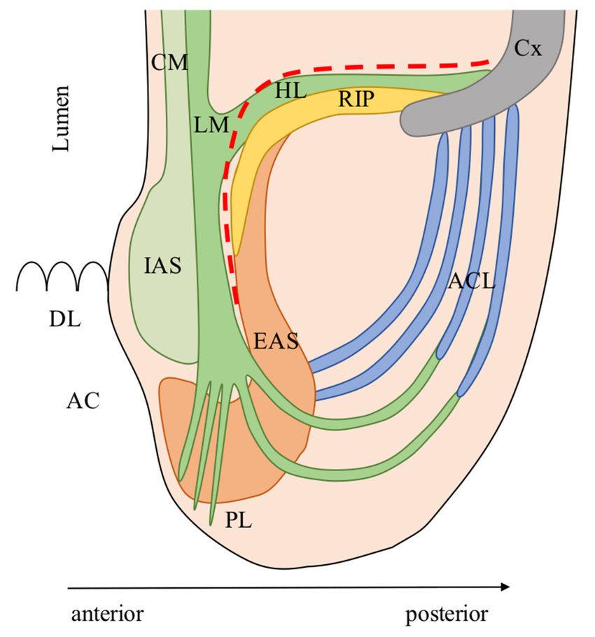 Posterior anatomy. AC: Anal canal; ACL: Anococcygeal ligament; Cx: Coccyx; CM: Circular muscle of the anal canal; EAS: External anal sphincter; HL: Hiatal ligament (sometimes referred as anococcygeal ligament by colorectal surgeons); IAS: Internal anal sphincter; LM: Longitudinal muscle of the anal canal; PL: Parks’ ligament; RIP: Raphe of iliococcygeus and pubococcygeus muscle; Red line: Dissection plane during intersphincteric resection.
Internal Anal Sphincter
- Thickened downward continuation of the inner circular muscle of the Rectum
- At anorectal junction, the outer longitudinal layer of the rectal muscle becomes fibroelastic and, together with some striated muscle fibres of Puborectalis, forms the Conjoint Longitudinal Coat
- Runs down between the two sphincters
- Strands from this sheet penetrate the Internal Sphincter and lower part of the External Sphincter
- Some reach the fat of the Ischioanal Fossa and peri-anal skin
- Some pass through the Internal Sphincter to the mucosa of the Anal Canal, esp. at the Pectinate Line
- Strands tethering the mucous membrane named the Mucosal Suspensory Ligament
- NB: Internal Anal Sphincter has NO bony attachments
Mucous Membrane
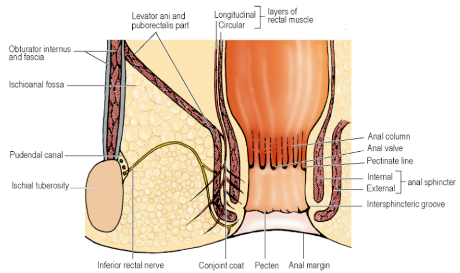
Anal Columns
- Located in the upper third of Anal Canal
- 6-10 longitudinal ridges
- Prominent in children
Anal Valves
- At lower ends of adjacent Anal Columns joined together by small horizontal folds
- NB: These are NOT palpable on rectal examination
- NB: Lie above a smooth surfaced area of the anal canal
Anal Sinuses
- Pockets formed above the Anal Valves
- Anal glands secret mucus into these sinuses
- Half of the Anal glands are submucosal; the rest penetrate the Internal Sphincter
- Infections in these glands cause anal abscesses and fistulae
Pectinate Line/Dentate Line
-
Level of Anal Valves
-
Below Pectinate Line:
- Pale, smooth surfaced area, the Pecten, extends down to the Intersphincteric Groove
- Below groove, area is truly cutaneous
- Histologically: Typical skin with keratinized stratified squamous epithelium, hair follicles, sebaceous glands and sweat glands
- Above the groove, the lining of the Pecten is non-keratinized stratified squamous epithelium, with no hair follicles, sebaceous glands or sweat glands
-
Anal column area – typical columnar intestinal cells and tubular glands
- Immediately above and below the Pectinate line, zone of variable often mixed epithelial structure
- Thus, no abrupt line of change from single-layered gut type to multi-layered pecten type
-
Above Dentate Line:
- Lymphatics drain to Inferior Mesenteric Lymph Nodes
- Receives autonomic innervation from the Inferior Hypogastric Plexus
- This region is sensitive to stretch
-
Below Dentate Line:
- Lymphatics drain to Superficial Inguinal Lymph Nodes
- Receives somatic innervation from branches of the Pudendal Nerve
- This region is sensitive to pain, temperature and touch
Anal Cushions
- Small submucous masses
- Comprises fibroelastic connective tissue, smooth muscle, dilated venous spaces and arteriovenous anastomoses
- Located at 3 (left lateral), 7 (right posterior) and 11 (right anterior) o’clock positions in upper anal canal
- Via their apposition, the Anal Cushions help the sphincter maintain water-tight closure of the Anal Canal
- Haemorrhoids – enlargement of Anal Cushions from excessive straining at stool
Development
- Lining of upper part of Anal Canal – derived from the Cloaca, i.e. endoderm
- Lining of lower part of Anal Canal – derived from Proctodeum or Anal Pit, i.e. ectoderm
- Dividing line between these territories considered to be at the Pectinate line
Blood Supply
- Superior Rectal Artery branches – supply upper end of Anal Canal
- Terminations lie within the Anal Columns
- Median Sacral Arteries – supply a small part of the muscular wall
- Inferior Rectal Arteries – supply lower end of Anal Canal including mucous membrane
- Vessels cross the Ischioanal Fossae
- Good anastomosis between the vessels within the walls
- Veins correspond to the above arteries
- Continuous with the Rectal Venous Plexuses
- Upper part of the canal and plexus drain via Superior Rectal and Inferior Mesenteric Veins to the Portal system
- Lower end drains to the Internal Iliac Veins through the Inferior and Middle Rectal Veins
- Anal Canal thus a site of porta-systemic anastomosis – union being in the region of the Anal Columns
Lymph Drainage
- Upper Anal Canal ⇒ Join lymphatics of Rectum
- Lower Anal Canal ⇒ Superficial Inguinal Lymph Nodes
Nerve Supply
-
Inferior Rectal branches of the [[Pudendal Nerve
- Supply the External Anal Sphincter
- Sensory supply from 1-2cm above the Pectinate Line downwards (where lining of Anal Canal is highly sensitive)
- Motor fibres originate from Onuf’s Nucleus – located mainly in anterior horn of S2 segment
- Also innervates the Sphincter Urethrae
-
Puborectalis and deep part of the External Anal Sphincter – high proportion of slow twitch fibres
- Function as tonic muscles
- Constant electromyographic activity even in sleep and light anaesthesia
-
Autonomic Nerves – pass to the Internal Anal Sphincter and Upper Part of the Anal Canal
-
Sympathetic fibres from the Pelvic Plexus:
- Pre-Ganglionic cell bodies in the first 2 lumbar segments of the cord
- Cause contraction of the Internal Anal Sphincter
- Pelvic Splanchnic (Parasympathetic) nerves relax it
-
Afferent fibres from upper end of the Anal Canal – carried by both sympathetic and parasympathetic nerves
Defecation
- Factors contributing to normal anal continence:
- Contraction of Puborectalis and External Anal Sphincter
- Maintenance of the angle between the Rectum and Anal Canal with abdominal pressure flattening the lower anterior rectal wall over the upper end of the canal
- Presence of Mucosal Cushions in the Anal Canal
- Internal Anal Sphincter
- Can only maintain continence if there is no distension (which causes relaxation of the sphincter)
- Rectum able to receive a certain amount of colonic content without any significant increase in pressure
- No specialized receptors in rectal wall
- Specialized receptors present in anal canal – gas, fluid and solid can be distinguished by the Cerebral Cortex
- Stretch receptors present in the Levator Ani and Peri-rectal tissue
- Increasing rectal pressure ⇒ Faeces enters upper anal canal ⇒ External Anal Sphincter contracts ⇒ Forces contents back into the Rectum
- If only gas enters, its presence can be tested by a slight conscious increase of abdominal pressure which will let it escape
- Defecation allowed to occur via release of cortical inhibition that developed during childhood training
- Abdominal pressure increases ⇒ Puborectalis relaxes ⇒ Anorectal angle straightens + Relaxation of External Anal Sphincter + Contraction of the lower Colon and Rectum (via parasympathetic supply)
- Incontinence:
- May follow damage to the External Anal Sphincter or Pudendal Nerve
- May occur in cerebral or spinal cord lesions with loss of cortical control
Ischioanal Fossa
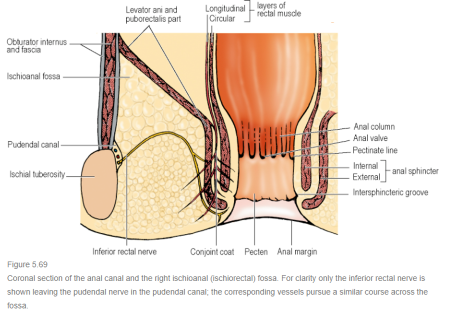
- Wedge-shaped space filled with fat lateral to the Anal Canal
- Base: Skin over the anal region of the Perineum
- Medial wall: External Anal Sphincter of Anal Canal and sloping Levator Ani muscles
- Lateral wall: Ischial Tuberosity below with Obturator Internus (covered by its fascia) above
- Apex of wedge = Where the medial and lateral walls meet – where the Levator Ani is attached to its tendinous origin over the Obturator Fascia
- Base of anterior boundary – posterior border of the Perineal Body and muscles of the Urogenital Diaphragm
- Posterior boundary – Sacrotuberous Ligament overlapped by the lower border of Gluteus Maximus
- Fat in lower part of the fossa adjacent to the skin – small lobules
- Fat in upper reaches of the fossa – large lobules
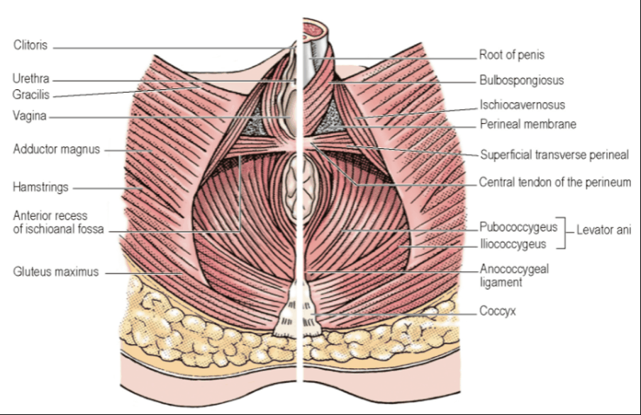
- Each fossa has an anterior recess that passes forwards above the Perineal Membrane
- Potentially as far as the posterior surface of the Body of the Pubis
- Recesses of two sides do not communicate across the midline
- NB: Thus, Ischiorectal Fossa extends anteriorly above the Urogenital Diaphragm
- Because the Ischiorectal Fossa extends forwards below the Levator Ani muscles which are attached to the Body of the Pubic Bone above the level of the Urogenital Diaphragm
- Posteriorly, the two fossae communicate with one another, low down through the fibrofatty tissue of the Retrosphincteric Space within the Anococcygeal Ligament
- Provides a horseshoe-shaped path for infection spread
[[Pudendal Canal
Of Alcock- Connective tissue tunnel in the lower lateral wall of the fossa
-
Overlies Obturator Internus and medial side of the Ischial Tuberosity
-
Canal formed by a splitting of the Obturator Fascia above the Falciform Process of the [[Sacrotuberous Ligament]]
-
Contains:
- Pudendal Nerve
- Internal Pudendal Vessels
-
The canal conducts from the Lesser Sciatic Notch to the Deep Perineal Pouch above the Perineal Membrane
-
Pudendal Nerve and Internal Pudendal Vessels leave the pelvis through the Greater Sciatic Foramen
- Passes beneath the lower border of Piriformis to reach the buttock
- Then turn and enters the Lesser Sciatic Foramen
- Vessels pass over the tip of the Ischial Spine and the nerve pass more medially over the Sacrospinous Ligament
-
Inferior Rectal Branches of the Pudendal Nerve:
- Run transversely across the Ischioanal Fossa from the Pudendal Canal towards the Anal Canal
- Course arches convexly upwards through fat towards apex and then down towards the canal
- Incisions to drain Ischiorectal abscesses do not usually interfere with them
- Supplies the External Anal Sphincter, mucous membrane of the lower Anal Canal and peri-anal skin
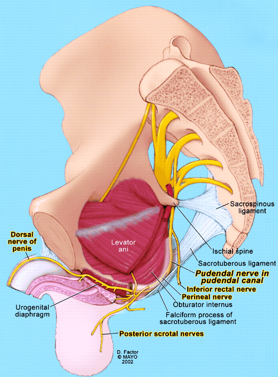
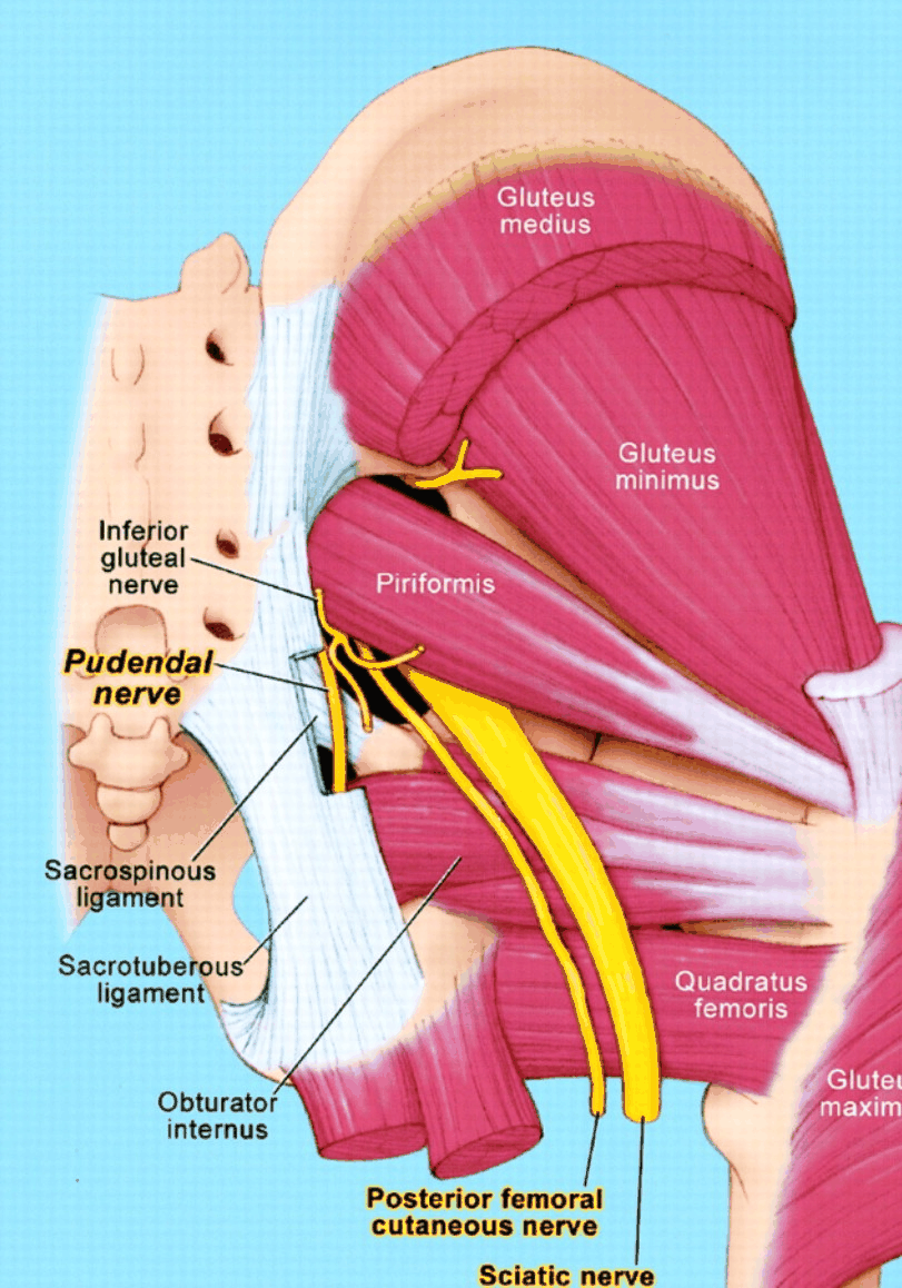
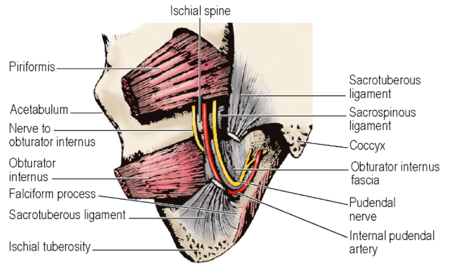 NB: Lateral wall of the Left Ischioanal Fossa from behind
-
At the front of the Ischioanal Fossa, the Posterior Scrotal (Labial) Nerves and Vessels (From the Pudendals) pass superficially into the urogenital region
-
At the back of the Ischioanal Fossa, the Perineal Branch of S4 Nerve and Perforating Cutaneous Nerve traverse the fossa
Perineal Body
- A.k.a. Central Tendon of the Perineum
- Midline fibromuscular mass
- Attached to the posterior border of the Perineal Membrane
- Lies between Anal Canal and Vagina or Bulb of the Penis
- Rectovaginal Septum blends into it above
- Muscles running into the Perineal Body include:
- External Anal Sphincter
- Pubovaginalis (Puboprostaticus) part of the Levator Ani
- Bulbospongiosus
- Superficial and Deep Transverse Perineal Muscles