External Oblique
- Origins
- 8 digitations, one from each of the lower 8 ribs
- The lower 4 interdigitate with the costal fibres of Latissimus Dorsi and the upper 4 interdigitate with Serratus Anterior
- Free posterior border extends from 12th rib to anterior half of the outer lip of the Iliac Crest
- 8 digitations, one from each of the lower 8 ribs
- Muscular fibres replaced by aponeurosis:
- Below a line joining ASIS to umbilicus
- Medial to a vertical line drawn from the tip of the 9th costal cartilage
- Aponeurotic fibres interdigitate with each other across the front of rectus abdominis along the whole length of the Linea Alba
- Free horizontal upper border of this aponeurosis extends from 5th rib to xiphisternum
- Only structure in the anterior sheath of the rectus muscle above the costal margin
- Lower border forms Inguinal Ligament (of Poupart) – between ASIS and pubic tubercle
- Edge rolled inwards – lateral part gives origin to part of the internal oblique and transversus abdominis muscles
- Fascia Lata of thigh attached to Inguinal Ligament
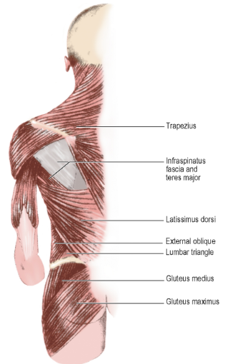
Lumbar Triangle of Petit (Inferior)
- Lateral: Posterior border of External Oblique
- Medial: Anterior border of Latissimus Dorsi
- Inferior: Iliac Crest
- Floor: Internal Oblique
- Site of inferior lumbar hernias
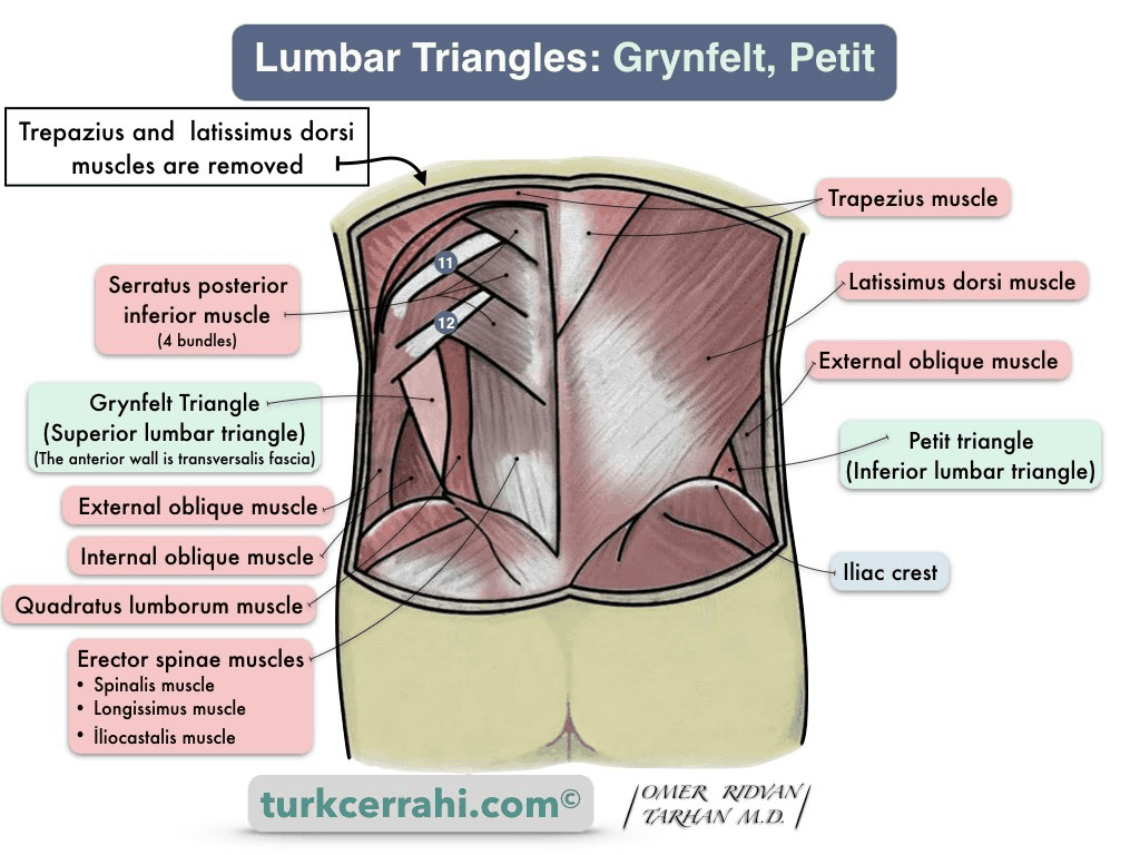
Lumbar Triangle of Grynfelt (superior)
- Lateral: Internal oblique
- Medial: Quadratus Lumborum
- Superior: 12th rib
- Floor: Transversalis fascia
- Site of superior lumbar hernias
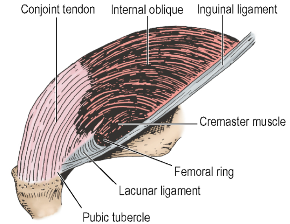
Internal Oblique
- Origin
- Whole length of Lumbar Fascia
- Anterior 2/3rds of the Iliac Crest
- Lateral 2/3rds of the Inguinal Ligament
- Insertion
- Ribs 10-12
- Course: From Lumbar Fascia, muscle fibres run upwards along costal margin
- Become aponeurotic at the tip of the 9th costal cartilage
- Below costal margin, aponeurosis splits around Rectus Abdominis – two layers rejoining at Linea Alba
- Free lower border – arches over the spermatic cord
- Lateral margin: Muscle fibres in front of the cord
- Medial margin: Tendinous fibres behind the cord
Arcuate Line (Semicircular Line of Douglas)
- Halfway between the Umbilicus and Pubic Symphysis
- Where the posterior layer ends in a curved free margin
- Below this point, aponeurosis passes wholly in front of the Rectus Abdominis to the linea alba
- May not be a well-defined line but a gradual diminution of aponeurotic fibres with increasing thickness of the Transversalis Fascia
Transversus Abdominis
- Origin
- Lateral 1/3rd of the Inguinal Ligament
- Anterior 2/3rds of the inner lip of the Iliac Crest
- Lumbar Fascia
- 11 and 12th Rib
- Inner aspects of the lower 6 Costal Cartilages (where it interdigitates with the diaphragm)
- Insertion
- Conjoint Tendon
- Xiphoid Process
- Linea Alba
- Pubic Crest
- Muscle fibres become aponeurotic and pass behind the Rectus Abdominis to fuse with the Internal Oblique aponeurosis in the Linea Alba
- Below Arcuate Line, aponeurosis passes wholly in front of the Rectus Abdominis
- In upper part of abdomen, the outer margin of the aponeurosis is more medial; muscular fibres lie behind the lateral part of the Rectus Abdominis
- Lower fibres of aponeurosis curve downwards and medially with those of the Internal Oblique as the Conjoint Tendon – inserting on the Pubic Crest and Pectineal Line
Iliopubic Tract
- Lower thickened part of the Transversalis Fascia between the Iliac Crest and the Pubis just above the Inguinal Ligament
- Band of fascial thickening of the transversalis fascia that lies on the posterior aspect of the inguinal region. The iliopubic tract runs deep and parallel to the inguinal ligament.
Conjoint Tendon
- The conjoint tendon is formed from the lower part of the common aponeurosis of the abdominal internal oblique muscle and the transversus abdominis muscle
- NB: Has a free inferolateral border
- NB: Attached to the superior pubic ramus
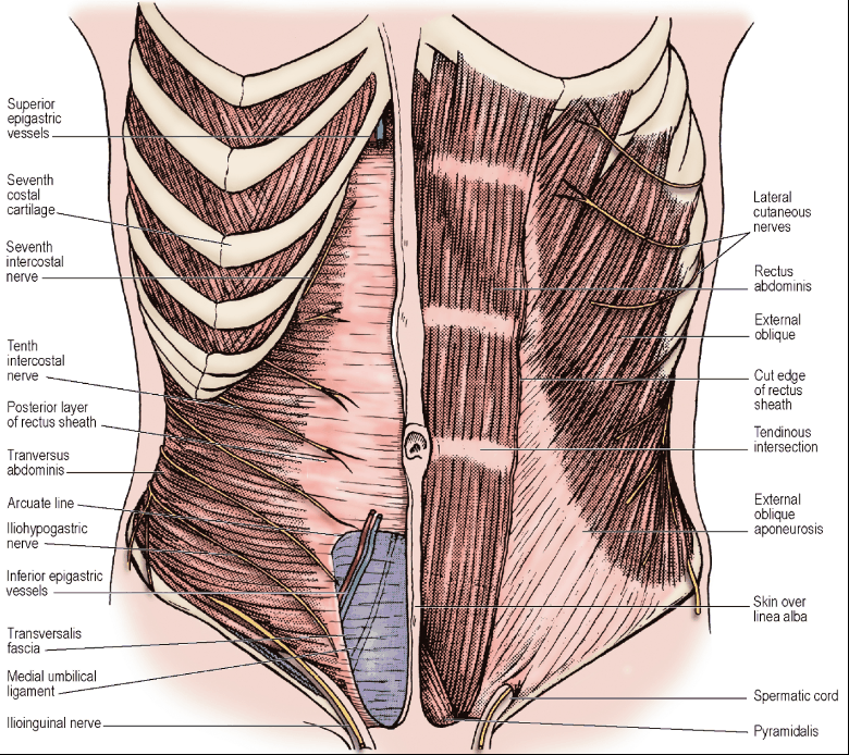
Rectus Abdominis
-
Origin: Two heads
- Medial head: In front of the Pubic Symphysis
- Lateral head: Upper border of the Pubic Crest
-
Upper parts of muscle separated from each other by the Linea Alba
-
Insertion: Front of the 5th-7th Costal Cartilages
- NB: Rectus Abdominis extends above the costal margin
-
Three tendinous intersections – at umbilicus, at xiphisternum and one between these two
- Intersections blend inseparably from the anterior layer of the Rectus Sheath
- Occupy only the superficial part of the muscle – do not penetrate to the posterior surface, thus, is not connected to the posterior layer of the sheath
-
Surgical Practice Point
- When performing a paramedian incision, to preserve the nerve supply to the whole of the rectus muscle – divide the Anterior Rectus Sheath and retract the Rectus muscle laterally instead of dividing it
- Keep the incision close to the midline
Pyramidalis Muscle
- Arises from body of Pubis and Pubic Symphysis between Rectus Abdominis and the Rectus Sheath - anterior to the recuts muscle
- Converges with its fellow into the Linea Alba ~4cm above its origin
Linea Alba
- Located between the two recti
- Formed from the fusion of all the aponeuroses that form the Rectus Sheath
- Attached to Xiphoid process above and Pubic Symphysis below
- Very narrow above the pubic symphysis – the two recti are in contact with one another behind it
- Umbilicus – defect in the Linea Alba through which the fetal umbilical vessels pass
Rectus Sheath
-
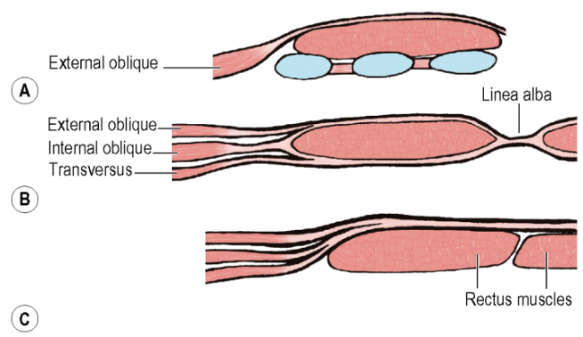
Internal Oblique aponeurosis splits into anterior and posterior layers to enclose the Rectus
-
External Oblique aponeurosis fuses with the anterior layer to the form the anterior layer of the Rectus Sheath
-
Transversus Abdominis aponeurosis fuses with the posterior layer to form the posterior layer of the Rectus Sheath
-
Below Arcuate Line, all three aponeuroses pass in front of the muscle
-
Posterior Layer of Rectus Sheath:
- Free lower margin – forms Arcuate Line (Semicircular Line of Douglas)
- Superior margin – attached to costal margin (7th, 8th and 9th costal cartilages)
-
Anterior Layer of Rectus Sheath:
- Above the costal margin – Consists of only the External Oblique aponeurosis
-
Rectus Sheath Contents:
- Rectus Abdominis
- Pyramidalis muscle
- Ends of the lower 6 thoracic nerves and their accompanying Posterior Intercostal vessels
- Superior Epigastric vessels
- Inferior Epigastric vessels
A = Above costal margin, B = Above Arcuate Line, C = Below Arcuate Line
Semilunar Line
- A shallow groove formed from the splitting of the Internal Oblique aponeurosis along the lateral border of the Rectus
- Curves up from the Pubic Tubercle to the costal margin at the tip of the 9th costal cartilage in the Transpyloric Plane