Overview: Lines and Regions
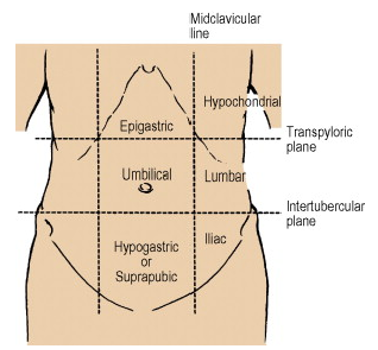
- Mid-clavicular line extends downwards to reach the Mid-Inguinal point, i.e. midway between Pubic Symphysis and ASIS
- Transpyloric Plane is midway between the Jugular Notch and the top of the Pubic Symphysis
- Subcostal plane is sometimes used – lower level
Transpyloric plane Landmarks
- Location
- Located between the Jugular Notch and the Pubic Symphysis
- Halfway between xiphisternum and umbilicus
- Passes through lower border of L1 Vertebra
- Cuts costal margin at the tip of the 9th Costal Cartilage
- At the lateral border of the Rectus Abdominis (Semilunar Line)
- Crosses
- L1
- Spinal cord ends at the Conus Medullaris
- Fundus of the Gallbladder
- Pylorus of the Stomach
- Duodenojejunal Flexure
- Head, Neck and Body of Pancreas
- Just above the attachment of the Transverse Mesocolon
- Supracolic Compartment lies above the plane
- Superior Mesenteric Artery leaves the Aorta
- Splenic Vein joins the Superior Mesenteric Vein to form the Portal Vein
- Hilum of each Kidney lies on this plane – the right just below and the left just above
- Hilum of Spleen
- L1
Trans-Tubercular Plane (L5) Landmarks
- Plane uniting the two tubercles of Iliac Crest
- Lies slightly posterior to the ASIS
- Fifth lumbar vertebra
- Confluence of Common Iliac Veins to form the Inferior Vena Cava (IVC) (2.5cm right of midline)
Anterolateral Abdominal Wall Muscles
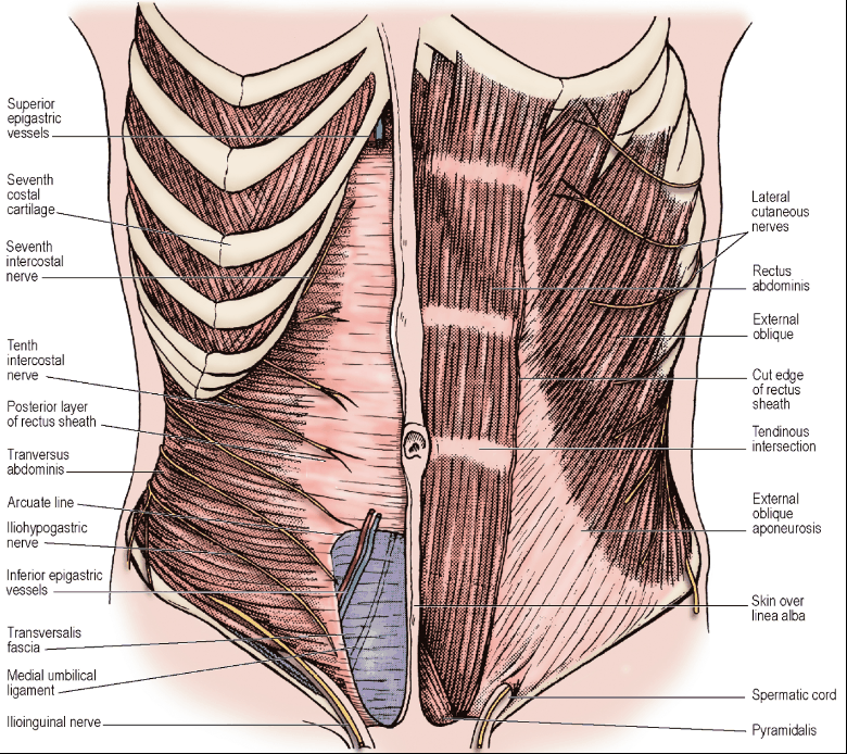
External Oblique
- Origins
- 8 digitations, one from each of the lower 8 ribs
- The lower 4 interdigitate with the costal fibres of Latissimus Dorsi and the upper 4 interdigitate with Serratus Anterior
- Free posterior border extends from 12th rib to anterior half of the outer lip of the Iliac Crest
- 8 digitations, one from each of the lower 8 ribs
- Muscular fibres replaced by aponeurosis:
- Below a line joining ASIS to umbilicus
- Medial to a vertical line drawn from the tip of the 9th costal cartilage
- Aponeurotic fibres interdigitate with each other across the front of rectus abdominis along the whole length of the Linea Alba
- Free horizontal upper border of this aponeurosis extends from 5th rib to xiphisternum
- Only structure in the anterior sheath of the rectus muscle above the costal margin
- Lower border forms Inguinal Ligament (of Poupart) – between ASIS and pubic tubercle
- Edge rolled inwards – lateral part gives origin to part of the internal oblique and transversus abdominis muscles
- Fascia Lata of thigh attached to Inguinal Ligament
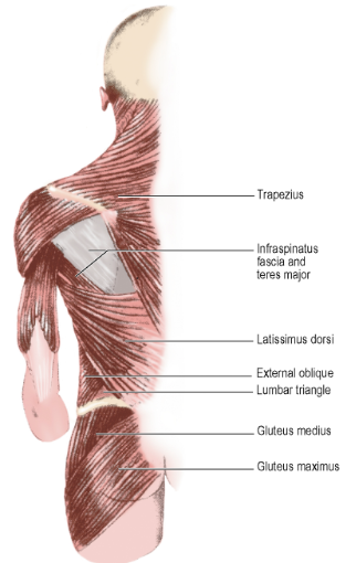
Lumbar Triangle of Petit (Inferior)
- Lateral: Posterior border of External Oblique
- Medial: Anterior border of Latissimus Dorsi
- Inferior: Iliac Crest
- Floor: Internal Oblique
- Site of inferior lumbar hernias
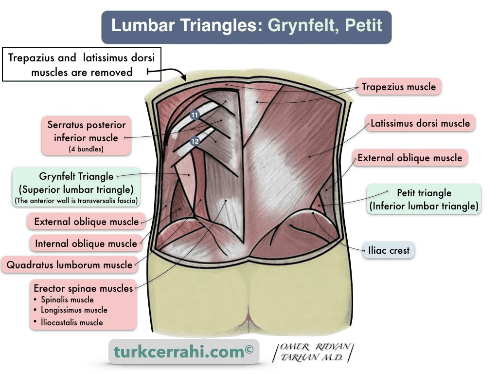
Lumbar Triangle of Grynfelt (superior)
- Lateral: Internal oblique
- Medial: Quadratus Lumborum
- Superior: 12th rib
- Floor: Transversalis fascia
- Site of superior lumbar hernias
Internal Oblique
- Origin
- Whole length of Lumbar Fascia
- Anterior 2/3rds of the Iliac Crest
- Lateral 2/3rds of the Inguinal Ligament
- Insertion
- Ribs 10-12
- Course: From Lumbar Fascia, muscle fibres run upwards along costal margin
- Become aponeurotic at the tip of the 9th costal cartilage
- Below costal margin, aponeurosis splits around Rectus Abdominis – two layers rejoining at Linea Alba
- Free lower border – arches over the spermatic cord
- Lateral margin: Muscle fibres in front of the cord
- Medial margin: Tendinous fibres behind the cord
Arcuate Line (Semicircular Line of Douglas)
- Halfway between the Umbilicus and Pubic Symphysis
- Where the posterior layer ends in a curved free margin
- Below this point, aponeurosis passes wholly in front of the Rectus Abdominis to the linea alba
- May not be a well-defined line but a gradual diminution of aponeurotic fibres with increasing thickness of the Transversalis Fascia
Transversus Abdominis
- Origin
- Lateral 1/3rd of the Inguinal Ligament
- Anterior 2/3rds of the inner lip of the Iliac Crest
- Lumbar Fascia
- 11 and 12th Rib
- Inner aspects of the lower 6 Costal Cartilages (where it interdigitates with the diaphragm)
- Insertion
- Conjoint Tendon
- Xiphoid Process
- Linea Alba
- Pubic Crest
- Muscle fibres become aponeurotic and pass behind the Rectus Abdominis to fuse with the Internal Oblique aponeurosis in the Linea Alba
- Below Arcuate Line, aponeurosis passes wholly in front of the Rectus Abdominis
- In upper part of abdomen, the outer margin of the aponeurosis is more medial; muscular fibres lie behind the lateral part of the Rectus Abdominis
- Lower fibres of aponeurosis curve downwards and medially with those of the Internal Oblique as the Conjoint Tendon – inserting on the Pubic Crest and Pectineal Line
Iliopubic Tract
- Lower thickened part of the Transversalis Fascia between the Iliac Crest and the Pubis just above the Inguinal Ligament
- Band of fascial thickening of the transversalis fascia that lies on the posterior aspect of the inguinal region. The iliopubic tract runs deep and parallel to the inguinal ligament.
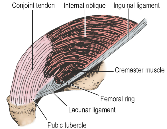
Conjoint Tendon
- The conjoint tendon is formed from the lower part of the common aponeurosis of the abdominal internal oblique muscle and the transversus abdominis muscle
- NB: Has a free inferolateral border
- NB: Attached to the superior pubic ramus
Rectus Abdominis
-
Origin: Two heads
- Medial head: In front of the Pubic Symphysis
- Lateral head: Upper border of the Pubic Crest
-
Upper parts of muscle separated from each other by the Linea Alba
-
Insertion: Front of the 5th-7th Costal Cartilages
- NB: Rectus Abdominis extends above the costal margin
-
Three tendinous intersections – at umbilicus, at xiphisternum and one between these two
- Intersections blend inseparably from the anterior layer of the Rectus Sheath
- Occupy only the superficial part of the muscle – do not penetrate to the posterior surface, thus, is not connected to the posterior layer of the sheath
-
Surgical Practice Point
- When performing a paramedian incision, to preserve the nerve supply to the whole of the rectus muscle – divide the Anterior Rectus Sheath and retract the Rectus muscle laterally instead of dividing it
- Keep the incision close to the midline
Pyramidalis Muscle
- Arises from body of Pubis and Pubic Symphysis between Rectus Abdominis and the Rectus Sheath
- Converges with its fellow into the Linea Alba ~4cm above its origin
Linea Alba
- Located between the two recti
- Formed from the fusion of all the aponeuroses that form the Rectus Sheath
- Attached to Xiphoid process above and Pubic Symphysis below
- Very narrow above the pubic symphysis – the two recti are in contact with one another behind it
- Umbilicus – defect in the Linea Alba through which the fetal umbilical vessels pass
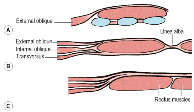
Rectus Sheath
-
Internal Oblique aponeurosis splits into anterior and posterior layers to enclose the Rectus
-
External Oblique aponeurosis fuses with the anterior layer to the form the anterior layer of the Rectus Sheath
-
Transversus Abdominis aponeurosis fuses with the posterior layer to form the posterior layer of the Rectus Sheath
-
Below Arcuate Line, all three aponeuroses pass in front of the muscle
-
Posterior Layer of Rectus Sheath:
- Free lower margin – forms Arcuate Line (Semicircular Line of Douglas)
- Superior margin – attached to costal margin (7th, 8th and 9th costal cartilages)
-
Anterior Layer of Rectus Sheath:
- Above the costal margin – Consists of only the External Oblique aponeurosis
-
Rectus Sheath Contents:
- Rectus Abdominis
- Pyramidalis muscle
- Ends of the lower 6 thoracic nerves and their accompanying Posterior Intercostal vessels
- Superior Epigastric vessels
- Inferior Epigastric vessels
A = Above costal margin, B = Above Arcuate Line, C = Below Arcuate Line
Semilunar Line
- A shallow groove formed from the splitting of the Internal Oblique aponeurosis along the lateral border of the Rectus
- Curves up from the Pubic Tubercle to the costal margin at the tip of the 9th costal cartilage in the Transpyloric Plane
Anterolateral nerves, vessels and lymph
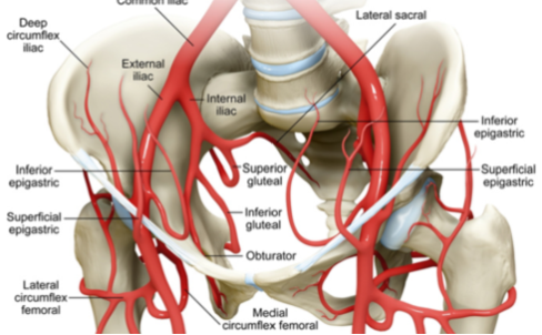
Superior Epigastric Artery
- Origin: Terminal branch of the Internal thoracic artery
- Course:
- Enters Rectus Sheath by passing between the sternal and highest costal fibres of the diaphragm
- Supplies: Rectus Abdominis
- Anastomoses within the Rectus muscle with the Inferior Epigastric Artery
- Veins drain to Internal Thoracic veins
Inferior Epigastric Artery
- Origin: External Iliac Artery at the Inguinal Ligament
- Course:
- Pass upwards behind the Conjoint Tendon
- Slips over the Arcuate Line to enter the Rectus Sheath
- Veins drain to External Iliac veins
Deep Circumflex Iliac Artery
- Origin: External Iliac Artery behind the Inguinal Ligament
- Course:
- Runs laterally towards the ASIS in a sheath formed by the Transversalis and Iliac Fasciae where they meet
- Continues along inner lip of Iliac Crest
- Pierces the Transversus Abdominis
- Travels within neurovascular plane
- Anastomose with branches of the Iliolumbar and Superior Gluteal Arteries
- At the ASIS, it gives an ascending branch that may be at risk in a gridiron incision
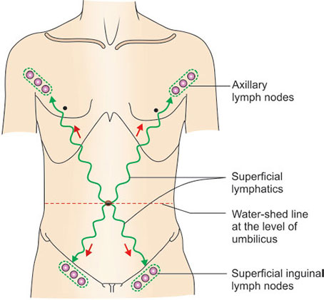
Lymph Drainage
- Superficial tissues – Drain in quadrants
- Pectoral group of Axillary Nodes above the umbilicus on each side
- Superficial Inguinal nodes below the umbilicus
- Deeper areas – Drain into vessels in the extraperitoneal tissues
- Above umbilicus, these pierce the diaphragm to reach the Mediastinal nodes
- Below umbilicus, they run to the External Iliac and Para-aortic nodes
Intercostal Nerves
-
T7 to T12
-
Course:
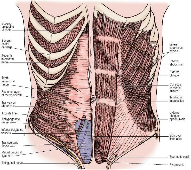
- Pass from intercostal spaces into abdo wall between the Internal Oblique and Transversus Abdominis muscles
- Travel within this neurovascular plane
- Before reaching sheath, they give off their Lateral Cutaneous branches
- Nerves pierce the Internal and External Oblique muscles to reach the skin
- Pierces the posterior layer of the Internal Oblique aponeurosis to enter the Rectus Sheath
- Proceed behind the Rectus to about its midline
- Pierce the Rectus to supply it
- Pass through the anterior layer of the Rectus Sheath to become the Anterior Cutaneous nerves
-
Surgical Practice Point
- Right Subcostal incision would sacrifice the 8th and 9th Intercostal Nerves but with little post-operative sequelae
- NB: Specifically, the Right 9th Intercostal Nerve is invariably cut in Kocher’s Subcostal Incision
- NB: 10th and, rarely, the 11th nerves may be divided only if the incision is extended laterally
- Right Subcostal incision would sacrifice the 8th and 9th Intercostal Nerves but with little post-operative sequelae
- NB: At the site of the McBurney’s Incision for Appendicectomy:
- The External Oblique is aponeurotic in most of its extent but fleshy fibres can be encountered laterally
- The Internal Oblique is almost entirely fleshy – it becomes aponeurotic just lateral to the Rectus Abdominis Muscle
- The Transversus Abdominis is half aponeurotic and half fleshy
Nerve Supply
- Rectus and External Oblique – supplied by the lower intercostal and subcostal nerves (T7 – T12)
- Internal Oblique and Transversus – supplied by same nerves with addition of Iliohypogastric and Ilioinguinal nerves (L1)
- L1 innervates the lowest fibres that continue medially as the Conjoint Tendon
- Pyramidalis – supplied by the Subcostal Nerve (T12)
Iliohypogastric Nerve
- Cross anterior surface of Quadratus Lumborum
- Derived from the 1st Lumbar Nerve (Anterior Ramus)
- Emerge from the lateral border of Psoas Major as a common stem with the Ilioinguinal Nerve which then divides or as separate nerves
- Lie posterior to the kidney
- Pierce the anterior layer of the Lumbar Fascia and pass anterior to it through Transversus Abdominis
- In the anterior abdominal wall, it lies in the neurovascular plane between the Internal Oblique and Transversus Abdominis muscles above the Iliac Crest
- Gives a Lateral Cutaneous Branch
- Pierces Oblique muscles above the Iliac Crest to supply skin of the upper part of the Buttock behind the area supplied by the Subcostal Nerve
- Continues downwards in the neurovascular plane and pierces Internal Oblique above the Anterior Superior Iliac Spine
- Pierces the External Oblique Aponeurosis 2.5cm above the Superficial Inguinal Ring and ends by supplying Suprapubic skin
Ilioinguinal Nerve
- The collateral branch of the Iliohypogastric Nerve
- Both derived from the 1st Lumbar Nerve (Anterior Ramus)
- No lateral cutaneous distribution
- Only has a terminal cutaneous distribution
- Cross the anterior surface of Quadratus Lumborum
- Lie posterior to the kidney
- Pierce the anterior layer of the Lumbar Fascia and pass anterior to it through Transversus Abdominis
- In the anterior abdominal wall, it lies in the neurovascular plane between the Internal Oblique and Transversus Abdominis muscles above the Iliac Crest
- Supplies their lower fibres which are inserted into the Conjoint Tendon
- Thus, important in maintaining integrity of Inguinal Canal
- Pierces the Internal Oblique and passes deep to the External Oblique
- Emerges on the front of the cord through the Superficial Inguinal Ring covered by External Spermatic Fascia
- Pierces the External Spermatic Fascia
- Distribution:
- Skin of the root of the Penis (or Clitoris)
- Anterior 1/3rd of the Scrotum (or Labium Majus/Mons Pubis)
- Small area of thigh just below the medial end of the Inguinal Ligament
Umbilicus
- Fibrous Cicatrix is a fusion of peritoneal and fibrous structures:
- Round Ligament of the Liver (Ligamentum Teres)
- Median Umbilical Ligament (Obliterated Urachus)
- 2 Medial Umbilical Ligaments (Obliterated Umbilical Arteries)
- Transversalis Fascia
- Umbilical Fascia around the Urachal Remnant
- Parietal Peritoneum
- In the fetus: transmits the Umbilical Arteries, Umbilical Vein, Urachus and the Vitelline Duct
- NB: Lies at the level between L3 and L4
- NB: Lies approximately midway between the Xiphoid and the Pubis
Urachus
- A channel between the bladder and the umbilicus where urine initially drains in the fetus in the 1st trimester.
- Channel usually seals off around the 12th week of gestation
- A small fibrous cord remains – called the Median Umbilical Ligament
- Types of pathology:
- Patent urachus – causes varying amounts of clear urine to leak at the umbilicus
- Urachal sinus – the urachus did not seal close to the umbilicus leading to a blind ending tract – can be asymptomatic or present with infection with abdo pain and discharge.
Vitelline/Omphalomesenteric Duct
- Communication between the embryonic yolk sac and its primitive midgut during early human development
- Usually obliterated during the 8th week of gestation as the placenta replaces the yolk sac as the primary source of fetal nutrition
- Meckel’s diverticulum develops when incomplete obliteration of the Vitelline Duct occurs between the 5th and 7th week of gestation
Peritoneal Folds
- Six peritoneal folds on the posterior surface of the anterior abdominal wall – 1 above and 5 below the umbilicus
Above the umbilicus:
- Falciform Ligament
- Consists of 2 adherent layers of peritoneum connecting the anterior surface of the liver to the supraumbilical part of the anterior abdominal wall just to the right of midline and to the inferior surface of the diaphragm
- Contains the Ligamentum Teres (Obliterated remains of the Left Umbilical Vein)
- During development, this vein carries blood from the placenta and bypasses the liver via the Ductus Venosus – a venous shunt joining the IVC on the cranial side of the liver.
- Inferior margin deviates to the right and is attached to the notch for this ligament on the inferior liver border
- In portal hypertension, the increase in venous pressure can cause re-canalisation of the embryological left umbilical vein within the Falciform ligament that takes blood to abdominal wall veins.
Below the umbilicus:
- Median Umbilical Fold
- Contains the Median Umbilical Ligament – the obliterated remains of the Urachus
- Medial Umbilical Fold (x2)
- Contains the Medial Umbilical Ligament – the obliterated remains of the Umbilical Artery
- Lateral Umbilical Fold (x2)
- Contains the Inferior Epigastric Vessels
- Although called umbilical folds, these don’t reach as far as the umbilicus
Posterior Abdominal Wall
Overview
- 5 x Lumbar vertebrae project forwards into abdo cavity due to Lumbar Lordosis
- Midline forward projection enhanced by IVC and Aorta which lie in front of vertebral bodies
- Deep paravertebral gutters lie on each side of convexity
- Floor of Paravertebral gutters:
- Lumbar Fascia
- Psoas Major
- Quadratus Lumborum
- Iliacus below the Iliac Crest
- Other parts of Posterior Abdo Wall:
- Left and Right Crura of Diaphragm
- Adjacent parts of Diaphragm
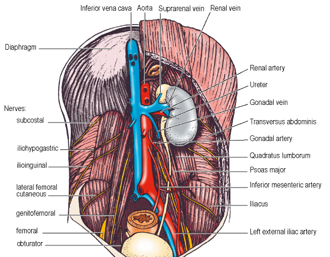
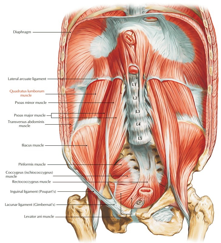
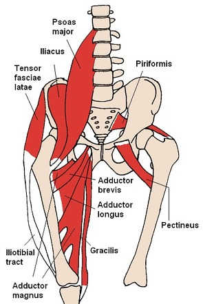
Psoas Major
- Lies in the gutter between bodies and transverse processes of lumbar vertebrae
- Origin:
- Discs above the 5 x Lumbar vertebrae (T12 – L5)
- Adjoining vertebral bodies
- Fibrous arches that span the concavities of the sides of the upper 4 vertebral bodies
- Medial ends of all Lumbar Transverse processes
- THUS, one continuous attachment from lower border of T12 to upper border of L5
- Course:
- Downwards along Pelvic Brim
- Passes beneath Inguinal Ligament into Thigh
- Insertion:
- Lesser Trochanter of Femur
- Relations:
- Lumbar Plexus embedded within muscle
- Genitofemoral Nerve and Femoral Nerves emerges from the front of the muscle
- Iliohypogastric, Ilioinguinal, Lateral Femoral Cutaneous, emerges from its lateral border
- Obturator Nerve and Lumbosacral Trunk emerge from its medial border
- 4 x Lumbar Arteries and veins pass backwards medial to the 4 arches and run laterally behind the muscle
- External Vertebral Venous Plexus behind it (in front of Transverse Processes)
- Psoas Fascia
- Invests surface of the muscle
- Attached to vertebral bodies, fibrous arches, and transverse processes
- Attached to Iliopubic eminence as muscle extends along pelvic brim
- NOT part of Lumbar Fascia
- Lateral edge blends with the anterior layer of Lumbar Fascia overlying Quadratus Lumborum
- Nerve Supply
- L1, L2 and L3 – mainly L2
- Actions:
- Hip flexion
- Lateral flexion of vertebra
- Flexion of trunk
Psoas Minor
- Present in 2/3 people
- Slender muscle lying on surface of Psoas Major
- Origin:
- T12 and L1 vertebrae
- Insertion:
- Iliopubic Eminence (as tendon flattens out to blend with Psoas Fascia)
- Nerve Supply:
- L1 Nerve
- Action:
- Weak flexion of lumbar spine
Iliacus
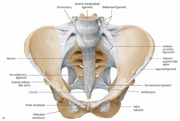
- Origin:
- Upper 2/3rds of the Iliac Fossa up to the Inner Lip of the Iliac Crest
- Anterior Sacroiliac Ligament
- Course:
- Fibres converge medially towards lateral margin of Psoas
- Pass out the Iliac Fossa beneath the lateral part of Inguinal Ligament
- Insertion:
- Psoas tendon below the Lesser Trochanter
- Iliac Fascia
- Strong fascia covering muscle
- Attached to bone at margins of the muscle and Inguinal Ligament
- Continuous with Psoas Fascia
- Forms a floor to the abdominal cavity
- Serves for the attachment of Parietal Peritoneum
- Nerve Supply:
- Femoral Nerve (L2, L3) in Iliac Fossa
- Action:
- Hip Flexion
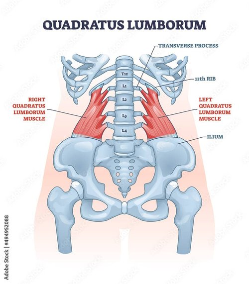
Quadratus Lumborum
- Lies edge to edge with Psoas Major medially and Transverse Abdominis laterally
- Lies in anterior compartment of Lumbar Fascia
- Represents the innermost of the 3 muscular layers of body wall, i.e. in series with Diaphragm and Transversus Thoracis muscle group
- Origins:
- Transverse process of L5
- Iliolumbar Ligament
- Iliac Crest
- Insertion:
- Transverse processes of L1 – L4 (Lateral to Psoas Major)
- Inferior border of medial half of 12th Rib
- Nerve Supply:
- T12 and L1-L3/L4 Lumbar Nerves
- Action:
- Prevent the diaphragm from elevating 12th rib during inspiration
- Lateral flexion of Lumbar Spine