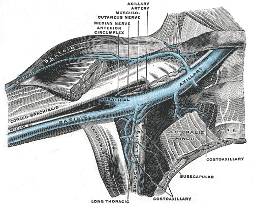
-
Commencement: Continuation of the Basilic Vein at the lower border of Teres Major- Terminates: Outer border of 1st Rib where it enters root of neck as the Subclavian Vein in front of Scalenus Anterior
-
Venae comitantes of Brachial Artery joins Axillary Vein near its commencement
-
Cephalic Vein drains into it above Pectoralis Minor
-
Divided into 3 parts based on relation to Pectoralis Minor – 1st part above, 2nd part behind and 3rd part below/lateral Relations:
-
Medial to Axillary Artery – which partly overlaps it anteriorly
-
Structure lying between Axillary Vein and Artery:
- Medial Pectoral Nerve
- Medial Cord of the Brachial Plexus
- Ulnar Nerve
- Medial Cutaneous Nerve of the Forearm
-
Lateral to the Medial Cutaneous Nerve of the Arm
-
Tributaries:
- 1st Part = Cephalic Vein
- 2nd Part = Same tributaries as 2nd part of the Artery, i.e. Lateral Thoracic and Thoracoacromial Veins
- 3rd Part = Same as 3rd part of Artery, i.e. Subscapular Vein, Anterior and Posterior Circumflex Humeral Vein
-
Other Veins
- Subscapular Veins
- Multiple and lie on posterior wall of axilla
- Lateral Thoracic Vein - **this can be used to find the vein in Axillary Lymph Node Dissection
- Subscapular Veins
-
Connected by the Thoracoepigastric Vein to the Superficial Epigastric Vein (tributary of the Great Saphenous Vein)
- Communication becomes prominent in cases of IVC obstruction
Relations
- Long thoracic nerve (nerve to serratus anterior) runs posteriorly in axilla on medial wall, deep to fascia on serratus anterior and runs behind axillary vein.
- Medial cutaneous nerve of the arm (from medial cord of the brachial plexus) runs down on the medial side of the axillary vein and supplies the skin over the front and medial side of the arm. This structure is at risk of damage during axillary dissection.
- Medial cutaneous nerve of the forearm runs between the axillary vein and artery and therefore should not be at risk during axillary dissection, given that axillary dissection should be confined to the axillary contents inferior and medial to the axillary vein.
- Thoracodorsal nerve (nerve to latissimus dorsi) runs behind the axillary vein and is initially posterior and medial to the subscapular artery.
- The 2 structures then run down the latissimus dorsi.
- Preserve these structures during axillary dissection to ensure neurovascular supply to the latissimus dorsi muscle which may be required at a later date for either rotation or free flap myocutaneous reconstruction.
- Nerve runs from medial to lateral to join the vein
- The lateral cutaneous branch of the second intercostal nerve originates from T1/2 and is named the intercostobrachial nerve.
- It pierces the Intercostalis externus and the Serratus anterior, crosses the axilla to the medial side of the arm, and joins with a filament from the medial brachial cutaneous nerve. It then pierces the fascia, and supplies the skin of the upper half of the medial and posterior part of the arm, communicating with the posterior brachial cutaneous branch of the radial nerve. At risk as lies below axillary vein and traverses plane of dissection.
- The medial pectoral nerve (medial anterior thoracic) arises from the medial cord of the brachial plexus and through it from the eighth cervical and first thoracic roots.
- It passes behind the first part of the axillary artery, curves forward between the axillary artery and vein, and unites in front of the artery with a filament from the lateral nerve. It then enters the deep surface of the pectoralis minor muscle, where it divides into a number of branches, which supply the muscle.