Overview of Axilla
- Location: The space between the upper arm and the side of the thorax
- Pyramidal shaped space
- Passage from the root of the neck to the upper limb through which nerves, blood vessels, and lymphatics travel to and from the arm
Boundaries of the Axilla
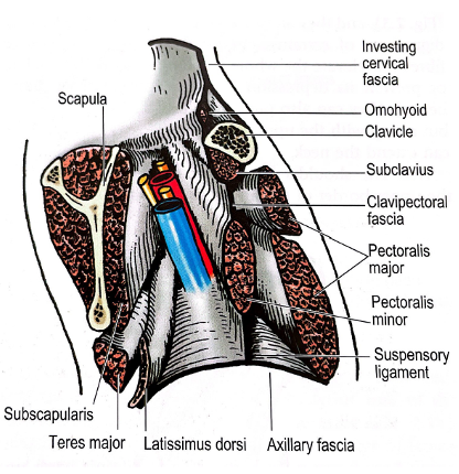
- Apex: Bounded by clavicle, upper border of scapula and outer border of 1st rib – channel of communication between axilla and posterior triangle
- Anterior and Posterior: Axillary folds
- Floor: Axillary fascia – extending from the anterior to the posterior axillary folds and from the fascia over serratus anterior to the deep fascia of the arm
- Suspensory ligament from lower border of pectoralis minor is attached to the fascial floor from above
- Anterior wall: Pectoralis major, pectoralis minor, subclavius, and clavipectoral fascia
- Posterior wall: Subscapularis and teres major – with tendon of Latissimus dorsi winding around teres major
- Medial wall: Upper part of serratus anterior
- Lateral wall: Coracobrachialis, Biceps, Bicipital Groove of Humerus
- Lower limit: Level of 4th rib
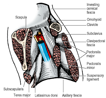 Vertical section of left axilla, looking laterally towards arm
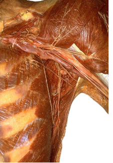
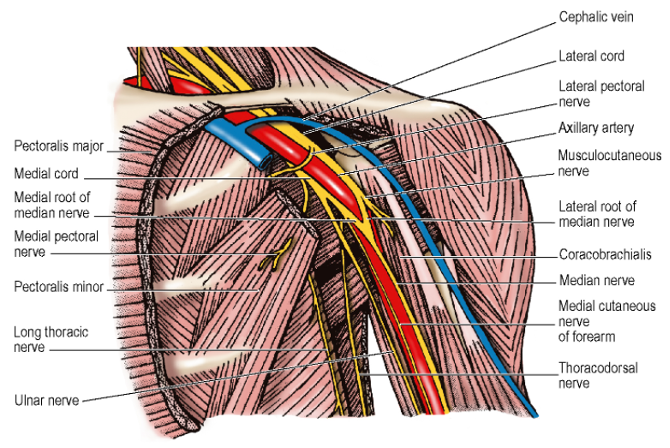 Left axilla from the front – pec major and minor and axillary vein removed
- Anterior and posterior walls converge laterally to the lips of the intertubercular groove of the humerus – in which lies the tendon of long head of biceps overlapped medially by coracobrachialis and tendon of short head of biceps.
Contents of Axilla
Contents Summary
- Vessels: Axillary artery, Axillary vein, Subscapular veins, Lateral Thoracic vein
- Nerves: Brachial plexus (cords) and branches, including Long Thoracic Nerve and Thoracodorsal nerve, also Intercostobrachial Nerve
- Lymphatics: Level 1, 2, and 3 Axillary lymph nodes
Axillary artery
-
Continuation of third part of subclavian artery
-
Commencement: Outer border of 1st rib
-
Course: Enters apex of axilla passing over first digitation of serratus anterior, behind midpoint of clavicle. At lower border of teres major, it becomes the brachial artery.
-
Divided into 3 parts by pectoralis minor, which crosses in front of it
-
First part
- One branch:
- Superior Thoracic Artery
- Runs forwards to supply both pectoral muscles
- Superior Thoracic Artery
- One branch:
-
Second part
- Two branches:
- Thoracoacromial Artery
- Skirts upper border of pectoralis minor ⇒ Pierce clavipectoral fascia – often separately by its 4 terminal branches – clavicular, deltoid, acromial and pectoral ⇒ Branches radiate away at right angles from each other in their respective directions
- Lateral thoracic artery
- Follows lower border of pectoralis minor ⇒ supplies branches to pectoralis and serratus anterior muscles
- In female: important contributor to blood supply of breast
- Thoracoacromial Artery
- Two branches:
-
Third part
- Three branches:
- Subscapular artery
- Largest branch – runs down posterior axillary wall ⇒ Gives off a dorsal branch – the Circumflex Scapular Artery which passes through posterior axilla wall between subscapularis and teres major, medial to long head of triceps and curves backwards round the lateral scapular border
- Distal to this large branch, the Subscapular Artery changes name to Thoracodorsal Artery⇒ Runs with nerve of same name into Latissimus Dorsi after giving 1-3 branches to Serratus Anterior
- Latter branches anastomose with Posterior Intercostal Arteries – thus, alternative source of blood supply to latissimus dorsi if subscapular or thoracodorsal artery has been occluded/divided higher up in axilla
- Thoracodorsal Nerve descends to lie anterior to the artery in a neurovascular bundle that enters latissimus dorsi close to its anterior edge.
- Anterior Circumflex Humeral Artery
- Runs deep to coracobrachialis and both heads of biceps
- Gives off ascending branch that runs up intertubercular sulcus and is an important blood supply to the humeral head
- Passes around surgical neck of humerus to anastomose with posterior circumflex humeral artery
- Posterior Circumflex Humeral Artery
- Passes through quadrangular space in posterior axillary wall, i.e. between subscapularis and teres major, lateral to long head of triceps and medial to the humerus
- Accompanied above by axillary nerve
- Supplies deltoid
- Gives branches to long and lateral heads of triceps and shoulder joint
- Anastomoses with profunda brachii artery
- Subscapular artery
- Three branches:
-
Axillary vein lies anteromedial to all parts of the artery
-
If arm at the side – artery has bold curve with convexity lateral. If arm abducted and externally rotated, e.g. during surgery, artery has straight course
-
Surface marking: A line from the middle of the clavicle to the groove behind coracobrachialis
-
Surgical approach: transverse incision below clavicle (pectoralis major split in line of its fibres) OR an incision along the deltopectoral groove (pectoralis major retracted away from deltoid) Tendon of pectoralis minor divided. Cords of brachial plexus and their branches safe guarded
Axillary Vein
-
Commencement: Continuation of the Basilic Vein at the lower border of Teres Major- Terminates: Outer border of 1st Rib where it enters root of neck as the Subclavian Vein in front of Scalenus Anterior
-
Venae comitantes of Brachial Artery joins Axillary Vein near its commencement
-
Cephalic Vein drains into it above Pectoralis Minor
-
Divided into 3 parts based on relation to Pectoralis Minor – 1st part above, 2nd part behind and 3rd part below/lateral Relations:
-
Medial to Axillary Artery – which partly overlaps it anteriorly
-
Structure lying between Axillary Vein and Artery:
- Medial Pectoral Nerve
- Medial Cord of the Brachial Plexus
- Ulnar Nerve
- Medial Cutaneous Nerve of the Forearm
-
Lateral to the Medial Cutaneous Nerve of the Arm
-
Tributaries:
- 1st Part = Cephalic Vein
- 2nd Part = Same tributaries as 2nd part of the Artery, i.e. Lateral Thoracic and Thoracoacromial Veins
- 3rd Part = Same as 3rd part of Artery, i.e. Subscapular Vein, Anterior and Posterior Circumflex Humeral Vein
-
Other Veins
- Subscapular Veins
- Multiple and lie on posterior wall of axilla
- Lateral Thoracic Vein
- Subscapular Veins
-
Connected by the Thoracoepigastric Vein to the Superficial Epigastric Vein (tributary of the Great Saphenous Vein)
- Communication becomes prominent in cases of IVC obstruction
Axillary Sheath
- Projection down from the prevertebral fascia in the neck
- Encloses axillary artery and cord of the brachial plexus
- Brachial plexus nerve block achieved by injecting local into this sheath
Brachial Plexus
Overview
Roots (5):
- Anterior rami of C5-8 and T1 (after they have given their segmental supply to the prevertebral and scalene muscles)
- 3 nerves are derived from the roots
Trunks (3): - Upper trunk = C5 and C6 - 1 nerve derived from upper trunk - Middle trunk = C7 - Lower trunk = C8 and T1
Divisions: - Each of the 3 trunks divides into an anterior and posterior divisions - 0 nerves derived from divisions
Cords: - Lateral cord = Upper two anterior divisions - 3 nerves derived from lateral cord - Medial cord = Anterior division of lower trunk - Medial cord frequently receives fibres from anterior ramus of C7 - 5 nerves derived from medial cord - Posterior cord = All three posterior divisions - 5 nerves derived from posterior cord
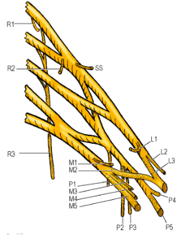
Relations
- Roots (5) lie behind scalenus anterior and emerge from between it and scalenus medius to form the trunks
- Trunks cross the lower part of the posterior triangle of the neck
- Divisions behind the clavicle
- Cords form at the outer border of the first rib
- Cords enter axilla above first part of axillary artery, approach and embrace its second part, and give off their branches around its third part
Variations
- Pre-fixed plexus: Significant contribution from anterior ramus of C4 and reduced contribution from T1
- Post-fixed plexus: Significant contribution from T2 and reduced contribution from C5
Branches from the Roots
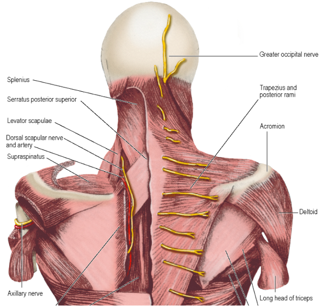
- Dorsal Scapular Nerve (nerve to the rhomboids)
- Arises from posterior aspect of C5
- Pierces Scalenus Medius ⇒ Courses downwards in front of Levator Scapulae ⇒ Lying on Serratus Posterior Superior
- Accompanied by dorsal scapular vessels
- Supplies both rhomboids and usually gives a branch to Levator scapulae
-
Nerve to Subclavius - Arises from the roots of C5 and C6 where they join to form the upper trunk
- Passes down in front of the trunks and subclavian vessels ⇒ Enter posterior surface of subclavius
- Frequently has a branch (accessory phrenic nerve) which connects with the phrenic nerve
- Alternative pathway for some fibres from C5 anterior ramus to reach the diaphragm
-
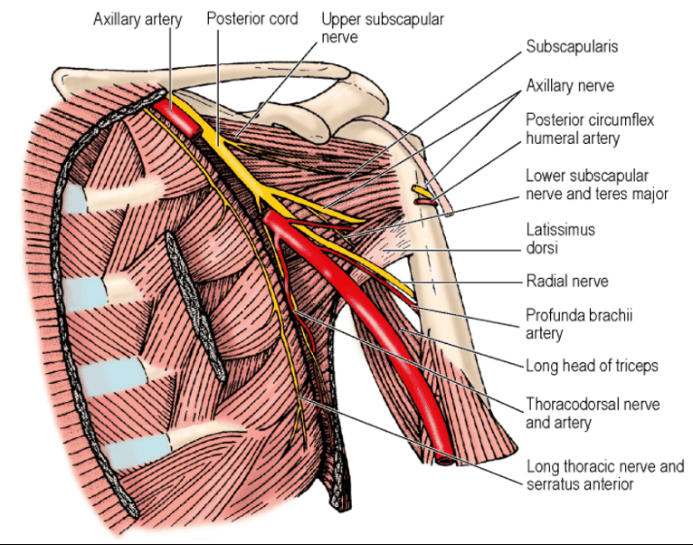
Long Thoracic Nerve (nerve to Serratus Anterior)
- Arises from posterior aspects of C5, C6 and C7
- Branches of C5 and C6 enter Scalenus Medius ⇒ Unites in the muscle ⇒ Emerge as single trunk ⇒ Pass down into axilla ⇒ Joined by branch from C7 (which descended in front of Scalenus Medius) on surface of Serratus Anterior (medial wall of axilla) ⇒ Passes down posterior to the Midaxillary Line, deep to fascia on Serratus Anterior
- NB: Runs behind the first part of the Axillary Artery
- Supplies serratus anterior segmentally
- In proximal Axilla, nerve runs superficial to the Serratus Anterior fascia and embedded within fatty tissue [WB]
- Penetrates the superficial fascia to lie on Serratus Anterior deep to the fascia at the level of 4th and 5th intercostal spaces
- Posterior to Lateral Thoracic perforating vessels
- Search for Long Thoracic Nerve begins high in Axilla – found embedded within axillary tissue lying approximately 1cm away from chest wall
- Thin layer of adventitial tissue enveloping the lymphatic tissue is incised laterally and parallel to Long Thoracic Nerve, thus freeing the nerve
- Nerve can then be pushed medially towards chest wall
Branches from the Trunks
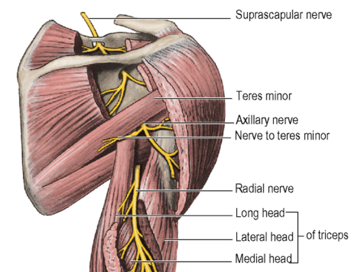
- Suprascapular Nerve
- Arises from the upper trunk in lower part of Posterior Triangle
- Passes backwards and laterally deep to border of Trapezius
- Passes through Suprascapular Foramen (beneath the Transverse Scapular Ligament)
- Supplies Supraspinatus
- Descends lateral to scapular spine with suprascapular vessels
- Supplies Infraspinatus
- Also supplies Shoulder and Acromioclavicular joints
Branches from the Cords
Branches from the Lateral Cord (3 branches)
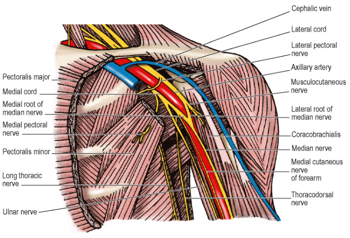
- Lateral Pectoral Nerve
- C5, C6 and C7
- Pierces clavipectoral fascia
- Supplies Pectoralis major
- Communicates across front of 1st part of Axillary artery with the Medial Pectoral Nerve
- Through this communication, supplies Pectoralis minor
- No cutaneous branch
- Musculocutaneous Nerve
- C5, C6 and C7
- Leaves lateral cord high in the axilla
- Runs obliquely downwards
- Passes through coracobrachialis
- Supplies: Coracobrachialis, Biceps brachii, Brachialis
- Becomes Lateral Cutaneous Nerve of the Forearm
- NB: Local anaesthetic injected through floor of axilla for brachial plexus nerve block may not affect Musculocutaneous nerve due to its high take off from the lateral cord
- Lateral Root of Median Nerve
- C5, C6 and C7
- Continuation of the Lateral Cord
- Joined by the Medial Root of the Median Nerve
- Two roots embrace the Axillary artery
- When arm pulled down to depress shoulder, the roots may compress the vessel
Branches from the Medial Cord (5 branches)
-
Medial Pectoral Nerve
- C8, T1
- Arises from medial cord behind 1st part of Axillary Artery
- Joined by a communication from the Lateral Pectoral Nerve
- Enters deep surface of Pectoralis Minor (supplying it)
- Perforates Pectoralis Minor
- Enters Pectoralis Major
- Ends by supplying lower costal fibres
- No cutaneous branches
-
Medial Root of the Median Nerve
- C8, T1
- Continuation of the Medial Cord
- Crosses Axillary artery to join Lateral Root of the Median Nerve
-
Medial Cutaneous Nerve of the Arm
- C8, T1
- Smallest branch
- Most medial branch
- Runs down medial side of Axillary Vein
- Supplies skin over the front and medial side of arm
-
Medial Cutaneous Nerve of the Forearm
- C8, T1
- Runs down between Axillary Artery and Axillary Vein in front of the Ulnar Nerve
- Supplies skin over the lower part of the arm and medial side of forearm
-
Ulnar Nerve
- C7, C8, T1
- Largest branch of Medial Cord
- Runs down between Axillary Artery and Axillary Vein
- The most posterior of the structures which run down the medial side of the flexor compartment of the arm
- May receive its C7 fibres as a branch from the Lateral Cord if these have not already passed to the Medial Cord from the anterior ramus of C7
Branches from the Posterior Cord (5 branches)
-
Upper Subscapular Nerve
- C5, C6
- Enters upper part of Subscapularis
-
Thoracodorsal Nerve
- C6, C7, C8
- Passes below Axillary Vein to enter Axilla [WB]
- Runs laterally to meet Subscapular Vessels – forms a triangular space over Subscapularis muscle
- Base of triangle: Axillary Vein
- Medial border: Thoracodorsal Nerve
- Lateral border: Subscapular vessels
- Runs down posterior axillary wall
- Crosses lower border of Teres major
- Enters deep surface of Latissimus dorsi – well forward, near the muscle border
- Comes from high up behind the Subscapular artery.
- As it descends to enter the muscle, it lies in front of the Subscapular artery (at this level, now called the Thoracodorsal artery)
- Thrown into prominence if humerus undergoes lateral rotation and abduction
- Lateral Thoracic Vein useful landmark for identification of Thoracodorsal nerve [WB]
- Thoracodorsal bundle located 1cm deep and medial to this vein
- Vein must be ligated and transected to expose the neurovascular bundle
-
Lower Subscapular Nerve
- C5, C6
- Supplies lower part of Subscapularis
- Ends in Teres major
-
Axillary Nerve
- C5, C6
- Supplies nothing in the axilla
- Runs backwards through the quadrangular space (bounded by subscapularis above, teres major below, long head of triceps medially, and surgical neck of humerus laterally)
- Passes just below capsule of shoulder joint with the Posterior Circumflex Humeral vessels below
- Emerges at the back of the axilla below Teres Minor
- After giving a branch to the shoulder joint, it divides into anterior and posterior branches
- Anterior branch: winds around behind the humerus in contact with the periosteum and enters deep surface of Deltoid supplying it
- Posterior branch: supplies Teres minor and Deltoid, then winds around posterior border of Deltoid to become the Upper Lateral Cutaneous Nerve of the Arm
-
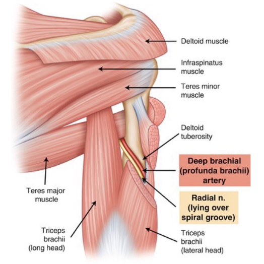
Radial Nerve
- C5, C6, C7, C8, T1
- Continuation of the Posterior Cord
- Crosses lower border of posterior axillary wall lying on the tendon of Latissimus dorsi
- Passes out of sight through Triangular Space below lower border of Latissimus dorsi tendon as it lies in front of Teres major, between Long Head of Triceps and Humerus
- Before disappearing, it gives branches to supply Long Head of Triceps AND the Medial Head of Triceps (a nerve which accompanies the Ulnar Nerve along the medial side of the arm) AND a cutaneous branch, the Posterior Cutaneous Nerve of the Arm supplying posterior surface of the arm
Axillary Lymph Nodes
-
General:
-
Varies between 20 and 30
-
Groups:
- Anterior or Pectoral Group
- Behind Pectoralis Major
- Along Lateral Thoracic Artery
- Along lower border of Pectoralis Minor
- Receive from the upper half of the trunk anteriorly and from the major part of the breast
- Posterior or Subscapular Group
- On posterior wall of axilla
- Along Subscapular artery
- Receive from the upper half of the trunk posteriorly and from the axillary tail of the breast
- Lateral Group
- Along medial side of Axillary vein
- Receive from the upper limb
- Central Group
- Receive lymph from above groups
- Apical Group
- Receives from all groups named above
- Drains into the Subclavian Lymph Trunk through apex of axilla into the Thoracic Duct or Right Lymphatic Duct or directly into the Jugulosubclavian venous junction in the neck
- A few efferents drain into the Supraclavicular (Inferior Deep Cervical) nodes
- Anterior or Pectoral Group
-
Alternatively, nodes described in terms of Levels
- Level 1 – Lateral to the lower border of Pectoralis minor
- Level 2 – Lie behind the muscle
- Level 3 – Lie medial to upper border of the muscle
Intercostobrachial Nerve
- The lateral cutaneous branch of the 2nd intercostal nerve
- Emerges from the 2nd intercostal space anterior to Long Thoracic Nerve and crosses axilla
- Supplies skin of axilla and variable extent on medial side of upper arm
- Often communicating with the Medial Cutaneous Nerve of the Arm
- May be in contact with level I lymph nodes and be at risk during node excision
- Thoracoepigastric Vein crosses the nerve vertically on its posterior aspect and aids identification
- Not infrequently: Lateral Cutaneous branch of the 3rd Intercostal Nerve also extends outwards to supply axilla skin
Pectoral Fascia
- Thin lamina of deep fascia that covers the anterior surface of Pectoralis Major
- Attachments:
- Medial: Sternum
- Above: Clavicle
- Lateral: Continuous with Axillary Fascia
- Forms floor of Retromammary space
- Gives origin to Platysma muscle from its upper part
Clavipectoral Fascia
- Strong fascial sheet deep to Pectoralis Major
- Upper part (a.k.a. Costocoracoid Membrane) – attached laterally to Coracoid Process and medially blends with the External Intercostal Membrane of the upper 2 spaces
- Splits above to enclose Subclavius
- Attached to edges of Subclavian groove on the undersurface of the Clavicle
- At lower border of Subclavius, the two layers fuse and form a well-developed band – the Costocoracoid Ligament
- Stretches from the knuckle of the Coracoid Process to the 1st Costochondral Junction
- From this ligament, the fascia stretches as a loosely felted membrane to the upper order of Pectoralis Minor
- Splits to enclose the muscle
- Below Pectoralis Minor, the fascia (a.k.a. Suspensory Ligament of the Axilla) extends downwards and attached to Axillary Fascia on the floor of the Axilla
- Its tension maintains the concavity of the Axilla
- Pierced by 4 structures:
- Two passing inwards, two passing outwards
- Inwards:
- Lymphatics from the Infraclavicular Nodes to the Apical Nodes of the Axilla
- Cephalic Vein
- Outwards: 3) Lateral Pectoral Nerve 4) Thoracoacromial Artery or its branches - Their corresponding veins join the Cephalic Vein anterior to the fascia
Potential Nerve Injuries in Axillary Dissection
-
Intercostobrachial nerve
- Lateral cutaneous branch of the 2nd intercostal nerve
- Emerges from the 2nd intercostal space anterior to the Long Thoracic Nerve and crosses the axilla
- Damage to this nerve could cause numbness of the skin overlying the axilla and a variable portion of the medial area of the upper arm.
- Key: Numbness or paraesthesia on the posterolateral aspect of upper arm
-
Thoracodorsal nerve
- Branch of the Posterior Cord of the Brachial Plexus
- Runs down the posterior axillary wall and crosses the lower border of Teres major
- Enters the deep surface of Latissimus dorsi
- Damage to this nerve would cause weakness of Latissimus dorsi leading to weakened extension, adduction and adduction of the ipsilateral arm.
- Key: Weak shoulder adduction
-
Long Thoracic nerve
- Arises from posterior aspects of C5, C6 and C7 nerve roots of the Brachial Plexus - Branches of C5 and C6 enter scalenus medius, unite in the muscle and emerge as a single trunk
- Branch from C7 descends anterior to scalenus medius and joins the above trunk on the surface of serratus anterior
- Then passes down posterior to the midaxillary line deep to the serratus anterior fascia
- Nerve supplies serratus anterior segmentally.
- Damage to this nerve causes winged scapula on the ipsilateral side.
Boundaries of Axillary Lymph Node Dissection
Medial Boundary
- Enter the Axilla
- Incising fascia 1cm below lateral edge of Pectoralis Major
- Ensures Medial Pectoral Nerve not injured
- Dissection continued cranially with simultaneous lateral retraction of axillary fat to help expose muscle edge
- Once lateral border of Pec Major exposed, it is then retracted anteriomedially to expose Pectoralis Minor underneath
- Dissection continued cranially freeing axillary fatty nodal tissue from lateral edge of Pectoralis Minor
Lateral Boundary
- Lateral border of Latissimus Dorsi
- Fascia overlying lateral edge of muscle incised parallel to muscle 1cm anterior to its edge
- Incision continued cranially until the tendon of the muscle or Axillary Vein is seen
- Thoracodorsal neurovascular bundle can be identified lying below the fascia overlying the Lat dorsi muscle (approximately 2cm medial to the lateral edge of muscle)
Superior Boundary
- Depends on purpose of axillary node dissection – breast cancer (level 2) or melanoma (level 3)
- Retracting Pec minor anteriomedially with elbow flexed, shoulder abducted allows exposure of Axillary Vein
- If inadequate exposure, Pec minor should be transected
- Dissection continued cranially until Axillary Vein identified behind medial edge of Pec Minor
- All fat and nodal tissue dissected off vein and swept laterally
- Superior dissection continued laterally along Axillary Vein, peeling down all lymphatic tissue and ligating all venous tributaries encountered until lateral extent of dissection reached
- Thoracodorsal Nerve emerges from beneath the Axillary Vein and courses laterally to meet the Thoracodorsal vessels
- Ligation of the Lateral Thoracic Vein is necessary to expose the Thoracodorsal neurovascular bundle
Inferior Boundary
- Where the angular veins drain into the Thoracodorsal vein
- When creating the medial and lateral boundaries, it frees the inferior boundary of axillary fat
Identification of Axillary Vein
Three main methods:
- Retrograde dissection by identifying angular veins draining into Thoracodorsal Vein
- Thoracodorsal vein then followed cranially until it enters Axillary Vein
- Latissimus dorsi tendon can be followed cranially also
- Palpation of Axillary Artery at apex of Axilla – vein lies anterior to the artery and posterior to the junction of the Clavicle and 1st Rib
- Retraction/Transection of Pectoralis Minor – located posterior to the medial edge of muscle
- Follow the lateral pectoral vein back
Related Muscles
- Pectoralis minor
- 3rd–5thrib
- Deep to pectoralis major to insert coracoid process
- n. lateral and medial pectoral nerves
- lateral from lateral cord brachial plexus
- medial from medial cord brachial plexus
- Pectoralis major
- Medial half clavicle, lateral half of sternum, 2nd to 6th costal cartilages and aponeurosis of external oblique
- Tendinous insertion into lateral lip intertubercular of humerus
- n. medial (C8) and lateral (C6,7) pectoral nerves
- paradoxically innervating the lateral and medial fibres respectively
- Serratus anterior
- Digitations of outer surface of 1-8th ribs
- Fibres pass backwards to insert along whole of medial border of scapula
- Holds scapula to chest wall
- n. long thoracic
- Subscapularis
- Costal surface of scapula
- Inserts lesser tuberosity of humerus
- Steadies head of humerus against glenoid cavity and assists in medial rotation
- n. upper and lower subscapular nerves from posterior cord which come off either side of thoracodorsal
- Teres Major
- Inferior angle of scapula
- Inserts into medial part of intertubercular groove of humerus
- n. lower subscapular nerves from posterior cord
- Latissimus Dorsi
- Lowermost three or four ribs, spinous processes of lower six thoracic vertebrae,
- lumbosacral vertebrae and iliac crest
- Spiral around lower border of teres major into intertubercular groove of humerus
- n. thoracodorsal nerve from posterior cord
Pectoralis Major
- Origins:
- Clavicular Head:
- Medial half of the anterior surface of the Clavicle
- Fibres lie on the manubrial part of the muscle running almost horizontally laterally (from which they are separate)
- Sternocostal Head:
- Lateral Half of the Anterior Surface of the Manubrium and Body of Sternum
- Upper 6 Costal Cartilages
- Aponeurosis of the External Oblique Muscle over the upper attachment of the Rectus Abdominis
- Clavicular Head:
- Insertions:
- Clavicular Head:
- Anterior lamina of the tendon into the Lateral Lip of the Intertubercular (Bicipital) Sulcus of the Humerus
- Sternocostal Head:
- Anterior lamina of the tendon into the Lateral Lip of the Intertubercular Sulcus behind (deep to) the Clavicular Fibres
- Lower Sternocostal fibres and abdominal fibres course upwards and laterally to be inserted progressively higher into the Posterior Lamina of the tendon
- Thus, producing the rounded appearance of the Anterior Axillary Fold
- The fibres which arise lowest inserted highest
- By a crescentic fold, blend with the capsule of the shoulder joint
- Clavicular Head:
- Lower medial part of the muscle is thinner
- At risk of perforation when a subpectoral pocket is created for insertion of a breast prosthesis
- Perforating branches of the Internal Thoracic Artery pierce the deep surface of the muscle at the Sternal edge
- At risk of being torn during subpectoral dissection
- Arterial Supply:
- Internal Thoracic Artery (perforating branches)
- Superior Thoracic Artery branches
- Lateral Thoracic Artery branches
- Pectoral branch of the Thoracoacromial Artery (Dominant branch)
- Nerve Supply:
- From the Brachial Plexus via the Lateral and Medial Pectoral Nerves
- Lateral Pectoral Nerve:
- Pierces the Clavipectoral Fascia medial to Pectoralis Minor
- Medial Pectoral Nerve:
- Pierce the Pectoralis Minor but may pass round its lateral border to reach Pectoralis Major
- Only muscle in the upper limb to be supplied by all 5 segments of the Brachial Plexus
- C5, C6 – Clavicular head
- C6-8, T1 – Sternocostal part
- Action:
- Adduction and Medial Rotation of the Arm
- Sternocostal part main adductors
- Flexion of the shoulder joint
- Via the Clavicular head
- NB: NOT a powerful flexor of the Gleno-humeral joint
- With upper limb fixed in abduction, it becomes an accessory muscle of inspiration, drawing the ribs upwards towards the Humerus
- Adduction and Medial Rotation of the Arm
- Testing:
- Clavicular Head: Arm abducted to 90 degrees or more, patient pushes arm forwards against resistance
- Sternocostal Head: Arm abducted to 60 degrees and then adducted against resistance
Pectoralis Minor
- Origin:
- 3rd, 4th and 5th ribs under cover of Pectoralis Major
- Insertion:
- Medial border and upper surface of the Coracoid Process of Scapula
- NOT the tip of the Coracoid Process – occupied by Biceps Brachii and Coracobrachialis
- Medial border and upper surface of the Coracoid Process of Scapula
- No great functional significance
- Muscle forms a tight band across the front of the Axillary neurovascular and lymphatic contents
- Division of its tendon allows for surgical clearance of the Axillary Lymph Nodes
- Nerve Supply:
- Both Pectoral Nerves (C6-C8)
- Action:
- Assists Serratus Anterior in Protraction of Scapula
- Keeps the Anterior (Glenoid) Angle in apposition with the chest wall as the Vertebral border is drawn forwards by Serratus Anterior
- Muscle elongated when Scapula rotates into full abduction of the arm
- Contraction assists gravity in restoring Scapula to rest position
- Assists Serratus Anterior in Protraction of Scapula
Subclavius
- Origin:
- Costochondral junction of the first rib
- Insertion:
- Subclavian groove on the inferior surface of the clavicle
- Nerve Supply:
- Nerve to Subclavius from the Upper Trunk of the Brachial Plexus (C5, C6)
- Action:
- Stabilizing the Clavicle in movements of the Pectoral Girdle
- May prevent the jagged ends of a fractured Clavicle from damaging the adjacent Subclavian Vein
Trapezius
- Origins:
- In the midline from the skull to lower thorax
- Extends from Medial third of the Superior Nuchal Line to the spine of C7 Vertebra
- Ligamentum nuchae between the External Occipital Protuberance and Vertebral Spine
- Extends along the spinous processes and Supraspinous Ligaments of all 12 Thoracic Vertebrae
- Insertions:
- Upper fibres: Posterior border of the Lateral third of the Clavicle at its posterior border
- Middle fibres: Medial border of the Acromion and the superior lip of the Crest of the Scapular Spine
- Fibres from the Lower 6 Thoracic Spines: Narrow recurved tendon into the medial end of the Scapular Spine
- Nerve Supply:
- Spinal part of the Spinal Accessory Nerve (C1-C5)
- Branches from the Cervical Plexus (C3 and C4)
- Usually only proprioceptive
- In some cases, may contain motor fibres also
- Nerves cross the Posterior Triangle to enter the deep surface of the Trapezius
- Accessory Nerve distinguished from the Cervical branches:
- Accessory Nerve emerges from within the substance of the Sternocleidomastoid
- Cervical nerves emerge from behind Sternocleidomastoid
- Action:
- Retract Scapula (all fibres)
- Upper and Lower fibres: Scapular rotation, tilting the glenoid upwards (essential component of shoulder abduction)
- Upper fibres: Elevate Acromion
- Lower fibres: Depress medial end of Spine
- Strongly assisted by lowest 4 digitations of Serratus Anterior
- Upper fibres: Elevate whole Scapula (shoulder shrug) or prevent depression (when carrying something heavy)
- Lateral flexion of neck
- Extend the neck (if acting with the upper fibres of the opposite side)
- Testing:
- Shoulder shrugged against resistance
Latissimus Dorsi
- Wide origin, narrow insertion
- Only muscle of the upper limb with a pelvic attachment (via the Lumbar Fascia)
- Origins:
- Spines of the lower 6 Thoracic Vertebrae
- Posterior layer of the Lumbar Fascia
- By which it is attached to the Lumbar and Sacral Vertebral Spines and posterior part of the Crest of Ilium
- Lateral to this: arises by muscular fibres from the Outer Lip of the Iliac Crest
- Course:
- Upper part: Runs horizontally covered medially by lower triangular part of Trapezius
- Passes over inferior angle of Scapula (from which a few fibres may arise)
- Middle part: Rubs obliquely upwards and outwards
- Lower part: Runs vertically upwards, reinforced by 4 slips from the lowest 4 ribs whose fibres of origin interdigitate with External Oblique fibres
- Lateral border forms a boundary of Lumbar Triangle
- Converges towards the Posterior Axillary Fold (forms the lower border)
- Sweeps spirally around the lower border of Teres Major (with some intermingling of fibres)
- Muscle then replaced by flattened, shiny, white tendon about 3cm wide
- Upper part: Runs horizontally covered medially by lower triangular part of Trapezius
- Insertion:
- Floor of the Intertubercular Sulcus
- Due to spiral turn around Teres Major – surfaces of muscle reversed at the tendon
- Glistening white tendon useful landmark to mark the lateral margin of the posterior wall of the Axilla during surgical dissection
- Occasionally, some muscle fibres from the edge of Latissimus Dorsi cross in front of the Axillary Vessels and nerves to blend with the tendon of Pectoralis Major forming a Muscular Axillary Arch
- Nerve Supply:
- Thoracodorsal Nerve (C6-C8) from Posterior Cord of the Brachial Plexus
- During its course down the posterior wall, it slopes forwards to enter the medial surface of the muscle just behind its anterior border in front of the Thoracodorsal Vessels
- Thoracodorsal Nerve (C6-C8) from Posterior Cord of the Brachial Plexus
- Actions:
- Extends shoulder joint
- Medial rotation of Humerus (e.g. folding the arms behind the back or scratching opposite Scapula)
- Adduction of Humerus (in combination with Pectoralis Major)
- When adducting upper limb from a position of abduction above the shoulder, plays important role in climbing
- Costal fibres can assist in deep inspiration
- Elevates the lower 4 ribs towards the fixed Humerus
- Rest of the muscle compresses the lower thorax in violent expiratory efforts, such as coughing or sneezing
- In spinal injury, it may move the pelvis and trunk
- Testing:
- Arm abducted to a right angle and then adducted, extended and medially rotated against resistance
Serratus Anterior
- Forms the medial wall of the Axilla
- Origins:
- Series of digitations from the upper 8 ribs
- First digitation arises from the 1st and 2nd ribs
- All other digitations arise from their corresponding ribs
- Lower 4 interdigitate with External Oblique
- Insertion:
- Costal (inner) surface of the Scapula
- 1st and 2nd digitations at the Superior Angle
- 3rd and 4th digitations as a thin sheet to the length of the Vertebral Border
- Lowest 4 digitations at the Inferior Angle
- Costal (inner) surface of the Scapula
- Muscle covered by a strong, well-developed fascia
- Nerve Supply:
- Long Thoracic Nerve (C5 – C7 roots of the Brachial Plexus)
- Nerve lies behind the Midaxillary Line (i.e. behind the lateral branches of the Intercostal Arteries) on the surface of the muscle
- Deep to the fascia
- Long Thoracic Nerve (C5 – C7 roots of the Brachial Plexus)
- Action:
- Protraction of the Scapula (whole muscle en masse) (e.g. during punching or pushing) – thus, elongating the upper limb
- Lower 4 digitations – powerfully assist Trapezius in rotating the Scapula Laterally and upwards in raising the arm above the level of the shoulder
- It is a more powerful rotator than the Trapezius
- In all positions of upper limb, it keeps the Vertebral Border of the Scapula in firm apposition with the chest wall
- Testing:
- With arm flexed and elbow extended, the outstretched hand is pushed against a wall
- Paralysis leads to ‘Winged Scapula’ where the Vertebral border becomes prominently raised off the Posterior chest wall