Overview
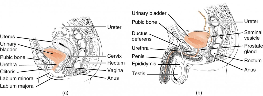
- Empty bladder situated entirely within pelvic cavity
- Empty bladder:
- Flattened 3-sided pyramid
- Sharp apex pointing forwards to the top of the pubic symphysis
- Triangular base facing backwards in front of the rectum or vagina
- Two inferolateral surfaces cradled by the anterior parts of the Levator Ani
- Neck – where Urethra opens
- Superior surface – on which small intestine and sigmoid colon or uterus lies
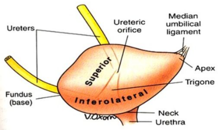
- Apex
- Attached to the remains of the Urachus, forming the Median Umbilical Ligament
- Runs up the midline of the anterior abdominal wall in the Median Umbilical Fold of the Peritoneum
- Attached to the remains of the Urachus, forming the Median Umbilical Ligament
- Base (Posterior Surface)
- Most lies below the level of the Rectovesical Pouch
- Only the uppermost portion is covered by peritoneum between the Vas Deferens on each side
- NB: Thus, can say the base is NOT covered by peritoneum
- Seminal Vesicles are applied to this surface
- Ureter enter in the upper outer corners
- In the female: The base has a firm connective tissue union with the anterior vaginal wall and upper part of the Uterine Cervix with no peritoneum intervening
- Inferolateral Surface
- Slopes downwards and medially to meet its opposite
- Lies against the front part of the Pelvic Diaphragm and Obturator Internus
- Retropubic Space of Retzius – the retroperitoneal space behind the pubic bones and pubic symphysis where the surfaces meet below the Apex
- Contains loose fatty tissue and the fibromuscular Pubovesical Ligaments that extend from the Bladder Neck to the Inferior Aspect of the Pubic Bones
- Neck
- Lowest part of the Bladder
- Where the base and inferolateral surfaces meet
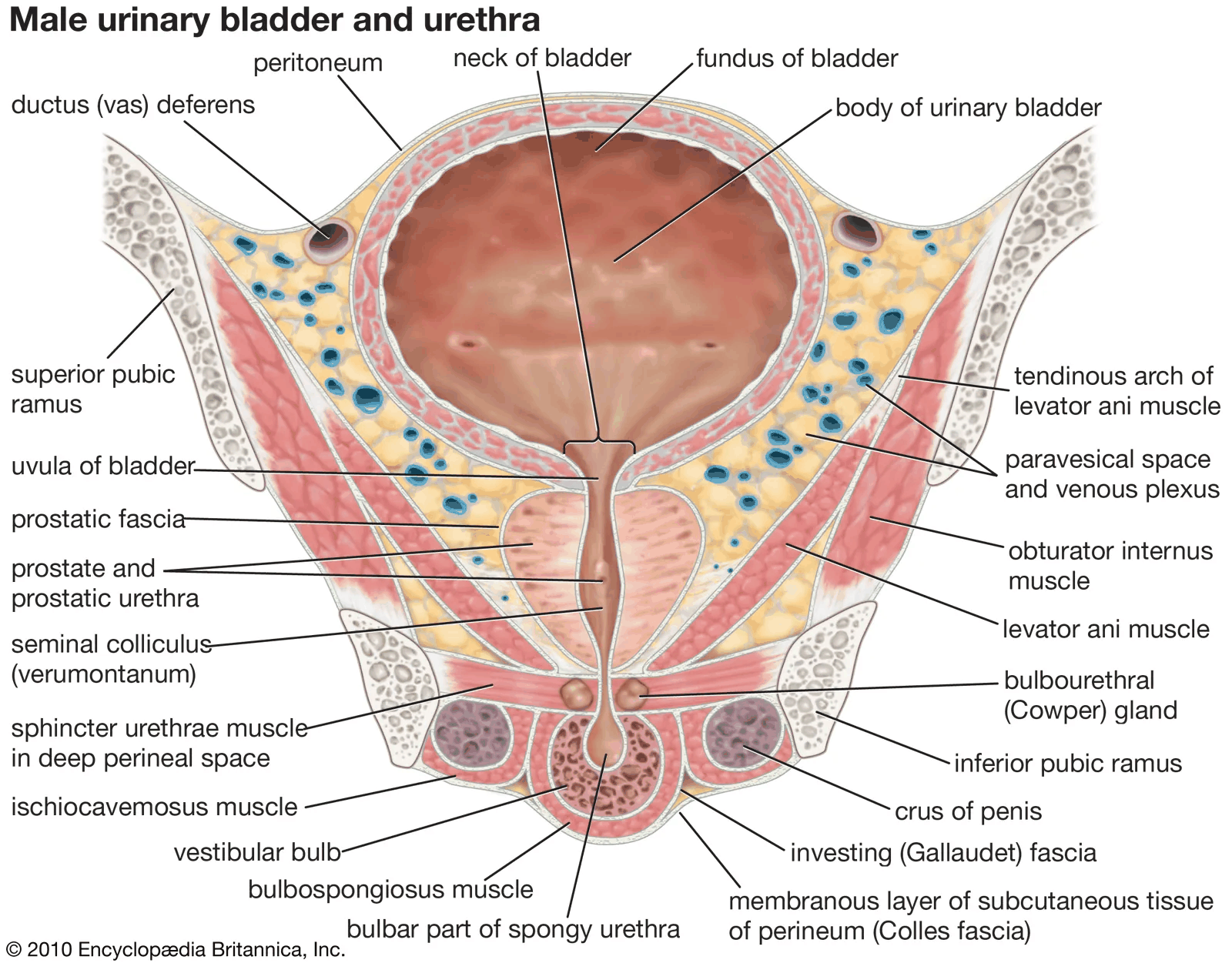
- Pierced by the Urethra at the Internal Urethral Orifice
- In the male: Lies against the upper surface or base of the Prostate
- In the female: Above the Urethra in the connective tissue of the anterior Vaginal Wall
- Superior Surface
- Covered by peritoneum (which sweeps upwards onto the anterior abdominal wall)
- Distending bladder strips peritoneum from behind the Rectus Abdominis, leaving the Transversalis Fascia on the back of the muscle
- Thus, distended bladder can be approached in the midline above the pubic symphysis by a cannula or scalpel without entering the peritoneal cavity
- In the male: At the posterior margin of this surface, the peritoneum continues onto the uppermost part of the base and is then continued backwards as the floor of the Rectovesical Pouch
- In the female: Reflected from a little in front of the posterior margin of this surface onto the under-surface of the Uterus
- Interior Appearance:
- Varies upon state of distension
- When collapse:
- Mucous membrane thick and thrown into folds
- When distended:
- Mucous membrane thin and smooth
- Trabeculae of muscle fibres can be seen through the mucous membrane
- These changes do not apply to the Trigone – varies little with state of distension
Trigone of Bladder
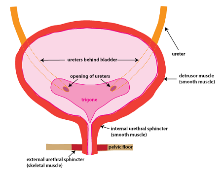
- Triangular area at the base of the bladder lying between the two Ureteral Orifices (above and laterally) and the Internal Urethral Orifice (centrally and below)
- In the empty bladder – these 3 openings are 2.5cm apart from each other
- When distended – ureteral orifices may be 5cm apart
- Ureteric Orifices connected by a transverse ridge – the Interureteric Bar
- Produced by the continuity of the longitudinal muscle of the two ureters across the bladder wall
- Ureters pierce the muscle and mucosal walls very obliquely
- Prevents reflux of urine
- Orifices closed by rising intravesical pressure
- Opens rhythmically in response to ureteric peristalsis each time a jet of urine is injected into the bladder (4-5 times per minute)
- Smooth walled
- Mucous membrane is relatively firmly adherent to the underlying muscle
- Fixed on-top of the prostate by the Urethra
- Trigone is the least mobile part of the Bladder
- In the male: Overlies the median part of the central zone of the Prostate
- After middle-age, the central zone may project above the internal urethral orifice as a rounded elevation, the Uvula of the Bladder
- In the female: Stabilized by the connective tissue surrounding the upper urethra at the front of the Vagina
Blood Supply
- Superior and Inferior Vesical Arteries
- Provide most of the arterial blood
- Small contributions to the lower part of the bladder from:
- Obturator Artery
- Inferior Gluteal Artery
- Uterine Artery
- Vaginal Arteries
- Veins do not follow arteries
- Form a plexus that converges on the Vesicoprostatic Plexus in the groove between Bladder and Prostate
- Drains backwards across the Pelvic Floor to the Internal Iliac Veins
- Similar plexus in females – communicates with veins in the base of the Broad Ligament
Lymph Drainage
- Mainly drain to External Iliac Nodes
- Some drain to Internal Iliac Nodes, including nodes in the Obturator Fossa
Nerve Supply
- Parasympathetic Fibres:
- Provide the main motor innervation of the Bladder
- Reach it via Pelvic Splanchnic Nerves
- Carries fibres for the sensation of normal bladder distension
- Conveyed in the spinal cord in the Gracile Tract
- NB: Carries sensory fibres from the Trigone of the Bladder
- Sympathetic Fibres:
- Come from L1 and L2 segments of the cord via the Superior and Inferior Hypogastric Plexuses
- Vasomotor for most of the Bladder
- Probably inhibitory to the Detrusor Muscle
- Motor to the Superficial Trigonal muscle and (in the male) the muscle of the bladder neck
- Bladder pain (e.g. from a stone) reaches the spinal cord (Lateral Spinothalamic Tract) by both Parasympathetic and Sympathetic pathways
Control of Micturition
- In general
- PNS → emptying
- SNS → storage
- Normal bladder emptying:
- Contraction of the Detrusor muscle and reciprocal relaxation of the external sphincter and pelvic floor (Levator Ani)
- Accumulation of urine distends Bladder wall with adjustment of tone (Accommodation) so that tension does not at first increase
- Later – increased tension stimulates stretch receptors
- Afferent impulses travel along Pelvic Splanchnic Nerves to sacral segments of the spinal cord
- Parasympathetic cell bodies stimulated and efferent impulses travel down the Pelvic Splanchnic Nerves to synapse with post-ganglionic cells within the bladder wall and cause contraction
- Autonomic stretch reflex with bladder control at the spinal level typical of infant
- Control by higher centres becomes superimposed on the spinal activity
- Bladder evacuation is assisted by voluntary contraction of abdominal muscles
- Cortical inhibitory centres in the Inferior Frontal Gyrus (on the medial surface of the cerebral hemisphere, some distance in front of the motor ‘Perineal’ area) with fibres passing to a Detrusor Motor Centre in the medial part of the Pontine Reticular Formation
- From there – Reticulospinal fibres run down the cord mixed with those of the Lateral Corticospinal Tract to the Sacral segments
- Skeletal Muscle of the Sphincter Urethrae (External Urethral Sphincter) – controlled by the Perineal Branch of the Pudendal Nerve
- Carries fibres predominantly from the Anterior Horn Cells of S2 Segment (Onuf’s Nucleus)
- Storage Centre in the lateral part of the Pontine Reticular Formation exerts central control on this nucleus
- During micturition, sphincter relaxes as the Detrusor muscle contracts
- In the female: the Pubovaginalis part of the Levator Ani assists the external sphincter at the end of micturition
- In spinal cord transection above the level of S2
- Afferent impulses indicating distension cannot reach consciousness
- Cortical control of the sacral reflex is lost
- Relaxation of the Sphincter Urethrae cannot be prevented
- Sacral centre intact, thus, the Bladder automatically empties when distended
- If sacral segments themselves are destroyed, the Detrusor muscle is paralysed and the bladder becomes abnormally distended until overflow incontinence occurs
Structure
- Smooth muscle of Bladder wall (Detrusor muscle) composed of interlacing network of fibres running in various directions
- Produce a trabeculated appearance (both externally and internally, beneath the mucous membrane)
- Exaggerated when muscular hypertrophy occurs as a result of progressive chronic obstruction to micturition
- Well supplied by Parasympathetic (Cholinergic) nerve fibres
- Trigone – possesses a superficial triangular layer of muscle (Superficial Trigonal Muscle) that is histologically and histochemically different from the rest of the Bladder (including the deep part of the Trigone)
- Extends into the proximal Urethra in both sexes
- Receives predominantly Sympathetic (Adrenergic) fibres
- Contraction of this muscle may help close the Ureteral Orifices
- In the male, at the Bladder Neck:
- Pre-Prostatic Sphincter: Circular smooth muscle fibres form a collar around the Internal Urethral Orifice
- Extends distally to surround the proximal part of the Prostatic Urethra (the Pre- prostatic part)
- Profusely supplied with Sympathetic (Adrenergic) fibres
- In the female, at the Bladder Neck:
- The muscle is arranged longitudinally and extends into the Urethral wall
- Muscle of the Bladder neck has nothing to do with urinary continence
- In the male: Acts to prevent seminal regurgitation into the bladder during ejaculation
- Mucous membrane:
- Thick
- Lax
- Lined by Transitional epithelium
- Glands usually absent (mucous in shed urine comes from Urethral glands)
- No muscularis mucosae
Development
- Endoderm of the Vesicourethral part of the Urogenital Sinus becomes the Bladder epithelium
- Surrounding mesenchyme forms the muscle and connective tissue
- Continued growth leads to incorporation of the lower ends of the mesodermal mesonephric ducts into the posterior part of the bladder – forming the Trigone
- The mesonephric ducts in the male end up at a lower level as the Ejaculatory Ducts entering the Urethra
- Allantois regresses to form a fibrous cord, the Urachus (median umbilical ligament)