Overview
- Length: 25cm
- Pelvic component forms about half of this length
- Points of narrowest calibre:
- Pelviureteric Junction
- Where it crosses the Pelvic Brim
- As it passes through the Bladder wall
- Adheres to the Peritoneum of the posterior abdominal wall when that membrane is stripped off the posterior abdominal wall
- Distinguished from vessels and nerves in the living body in that it is whitish, non-pulsatile cord which shows peristaltic activity when gently pinched with forceps.
Course and Relations
- Course:
- Pass down on Psoas Major under cover of Peritoneum
- Crosses in front of the Genitofemoral Nerve
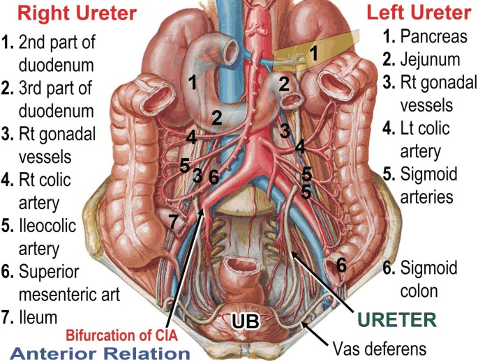
- Crossed anteriorly by the Gonadal Vessels
- On the Right:
- Upper part posterior to the 3rd part of the Duodenum
- Lower down – crossed anteriorly by the Right Colic and Ileocolic Vessels and Root of Small Bowel Mesentery
- On the Left:
- Lateral to the Inferior Mesenteric Vessels
- Crossed anteriorly by the Left Colic Vessels
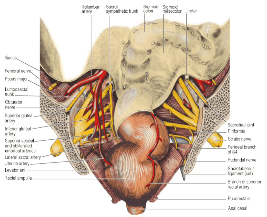
- At the Pelvic Brim, crossed anteriorly by the Apex of the Sigmoid Mesocolon
- Leaves Psoas Muscle at the bifurcation of the Common Iliac Artery over the Sacroiliac Joint
- Passes into the Pelvis
- Runs over the External Iliac Artery and Vein
- Runs down the side wall of the pelvis in front of the Internal Iliac Artery (Behind the Ovary)
- In order from above downwards, it crosses the:
- Obturator Nerve
- Obliterated Umbilical (Superior Vesical) Artery
- Obturator Artery
- Obturator Vein
- Turns forwards and medially above pelvic floor
- Enters base of Bladder at its upper lateral angles of the Trigone
- In the male: the Vas Deferens crosses above the Ureter and then runs down medial to the Ureter
- Upper end of Seminal Vesicles usually lies just below the point where the Ureter enters the Bladder Wall
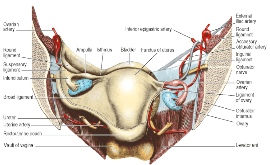
- In the female: Ureter lies in the base of the Broad Ligament where it is crossed above by the Uterine Artery
- Under the Broad Ligament, the Ureter penetrates the condensed tissue that forms the Lateral Cervical Ligament
- Crosses the Lateral Vaginal Fornix 1-2cm from the Cervix before entering the Bladder in front of the Fornix
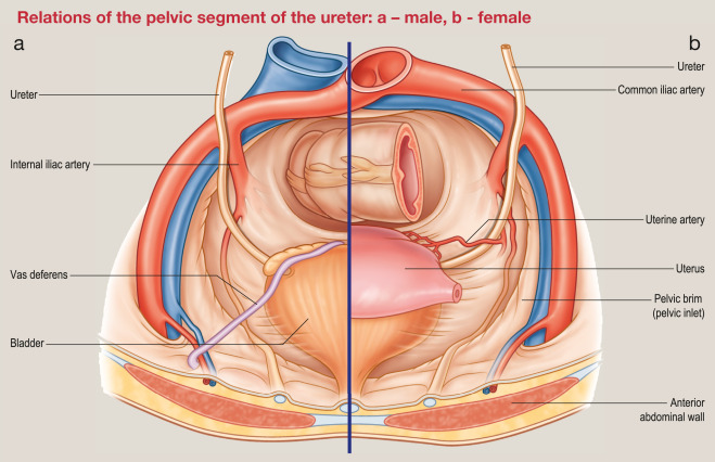
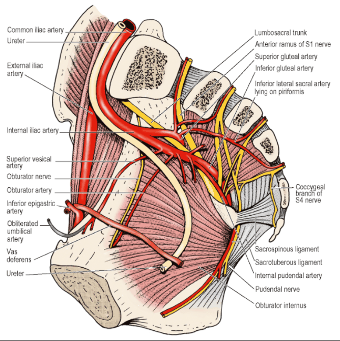
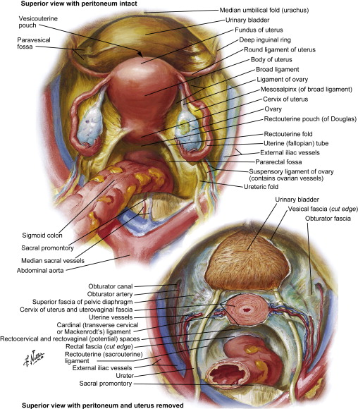
- In the male: the Vas Deferens crosses above the Ureter and then runs down medial to the Ureter
-
Locating the ureter intra-operatively:
- Over the bifurcation of the Common Iliac Artery over the Sacroiliac Joint
- At the apex of the sigmoid mesentry
- Chase the gonadal vessels as they will cross over the ureter
-
Surface markings:
- Anterior abdominal wall – marked from the tip of the 9th costal cartilage to the birfucation of the Common Iliac Artery
-
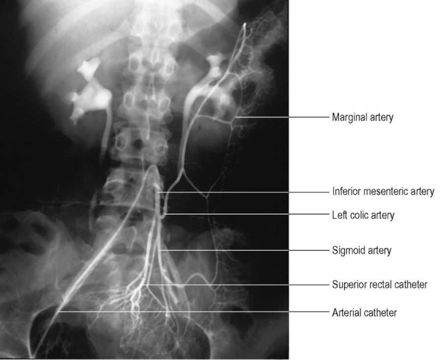
Line of projection of the Ureter on a radiograph:
- Lies medial to the tips of the transverse processes of the Lumbar Vertebrae
- Crosses Pelvic Brim at the Sacroiliac Joint
- From here, pelvic shadow passes to the Ischial Spine
- Then, foreshortened, to the Pubic Tubercle
Blood Supply
- Upper end: Ureteric branch of the Renal Artery
- Middle section: Abdominal Aorta, Gonadal, Common Iliac and Internal Iliac Arteries
- Lower end: Inferior and Superior Vesical and Uterine Arteries
- All vessels form good anastomosis with each other in the Adventitia of the Ureter forming longitudinal channels
- Blood supply is endangered if Ureter stripped clean of its surrounding tissue
Lymphatic Drainage
- Run back alongside the arteries
- Abdominal portion of Ureter ⇒ Drain to Para-aortic Nodes
- Pelvic portion of Ureter ⇒ Drain to Common Iliac and Internal Iliac Nodes
Nerve Supply
- Sympathetic fibres: From T10-L1 segments of the spinal cord via the Coeliac and Hypogastric Plexuses
- Parasympathetic fibres: From the Pelvic Splanchnic Nerves
- Functional significance of these innervations is unclear
- Intact innervation of Renal Pelvis and Ureter not necessary for the initiation or propagation of peristalsis from the Calyceal Pacemakers
- No ganglion cells in or on the Ureter
- Pain fibres – accompany sympathetic nerves
Structure
- Smooth muscle
- Lined internally by mucous membrane
- Lax
- Lined by transitional epithelium
- No muscularis mucosae
- Inner longitudinal, middle circular, and outer longitudinal
- More accurate to consider the muscle as a single coat with fibres running in many different directions
Development
- Mesodermal origin
- Derived by a process of budding from the caudal end of the Mesonephric Duct
- Upper end divides into 2 or 3 (the Major Calyces of the Renal Pelvis)
- Further subdivisions produce the Minor Calyces and Collecting Tubules
- Low division of the Ureteric Bud produces the Double Ureter