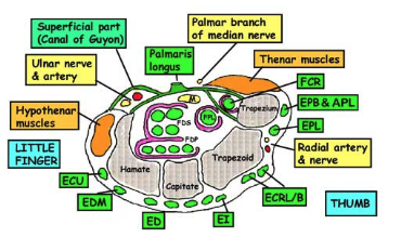
Overview
- Passageway on the palmar side of the wrist that connects the forearm to the hand.
- The tunnel is bounded by the bones of the wrist and flexor retinaculum from connective tissue
Carpal Bones
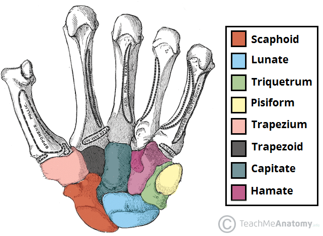 Some Lovers Try Positions That They Can’t Handle
Boundaries
- Superficial: Flexor retinaculum
- Flexor retinaculum:
- Medial attachment – Hook of Hamate and Pisiform
- Lateral attachment – Scaphoid tubercle and Trapezium
- Deep: Carpal bones
- Medial/Ulnar Border: Lateral surface of Hamate
- Lateral/Radial Border: Medial surface of Trapezium
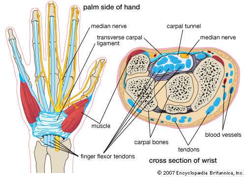
Contents
- Long flexor tendons of the fingers and thumb (9 tendons in total)
- Flexor Digitorum Superficialis (FDS) tendons lie in 2 separate rows – middle and ring finger tendons anterior to index and little finger tendons
- Flexor Digitorum Profundus (FDP) tendons lie in one row deeply with only tendon to index finger separate from the others
- All 8 tendons of FDS and FDP share common flexor sheath
- Sheath does not invest them completely but is reflected from their radial sides where arterial supply gain access – it is as if the tendons have been invaginated into the sheath from the radial side
- All 8 tendons of FDS and FDP share common flexor sheath
- Tendon of Flexor Pollicis Longus (FPL) lies in its own synovial sheath
- Median Nerve
- Passes deep to flexor retinaculum between FDS to the middle finger and Flexor Carpi Radialis tendon
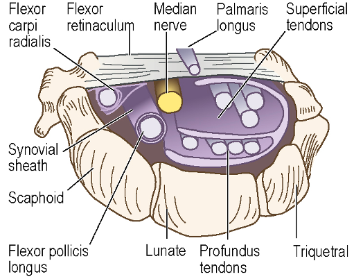 Figure: Left carpal tunnel, looking distally towards the palm
- Flexor Carpi Radialis (FCR), Flexor Carpi Ulnaris (FCU) and Palmaris Longus (PL) tendons travel outside the carpal tunnel but in close contact with it
- FCR - At lateral end of tunnel, enclosed in its own synovial sheath, the tendon runs in the groove within the groove on the Trapezium
- FCU – Tendon inserts into pisiform
- PL – Continues with palmar fascia and inserts into flexor retinaculum
Nerves at Risk During Carpal Tunnel Release
- Palmar Branch of Median Nerve
- Enters palm superficial to flexor retinaculum
- Recurrent Branch of Median Nerve
- Comes off median nerve distal to flexor retinaculum
- Curls proximally around distal border of flexor retinaculum to supply thenar muscles
Carpal Tunnel Syndrome
- Symptoms:
- Impaired sensation over 3 and a half digits on the lateral/thumb side of the hand
- In late cases: wasting and weakness of the thenar muscles
- NOTE: No sensory loss over thenar eminence ⇒ this area supplied by palmar branch of the median nerve which enters the palm superficial to the flexor retinaculum ⇒ escapes compression
- Must be distinguished from median nerve injury at a higher level
- If higher level ⇒ Palmar cutaneous branch affected and weakness of relevant flexor muscles, e.g. FPL, becomes notable
Carpal Tunnel Release Superficial Landmarks
- Carpal Tunnel Release
- Incision site: The intersection of the Kaplan cardinal line and the radial border of the 4th ray ending at the wrist crease
- Kaplan cardinal line = line drawn from the apex of the 1st web space (between thumb and index finger) toward the ulnar side of the hand, parallel with the proximal palmar crease.