Section: Skin and soft tissue Sub-section: Peripheral nerve issues Curriculum: Curriculum, page 55
Carpal Tunnel anatomy
Clinical
- Constellation of symptoms (paraesthesia, numbness, weakness of thenar muscles) resulting from Median Nerve compression at the wrist (? inflammatory, oedema, amyloid or collagen deposition)
- Tingling and numbness in radial 3 ½ digits.
- Deep aching pain in hand going up forearm – hallmark – wakes from sleep
- Thenar muscle atrophy late
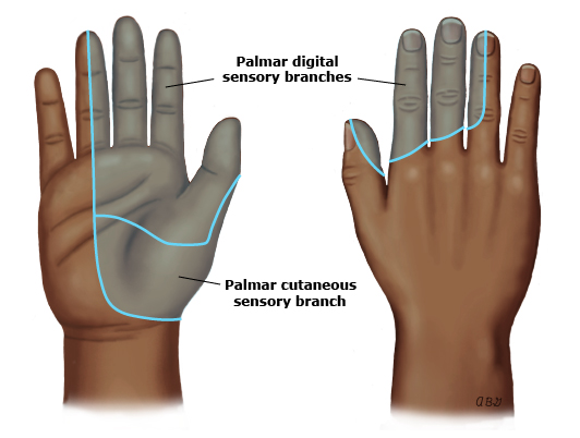
- NB: No sensory loss over thenar eminence ⇒ Area supplied by Palmar branch of the Median nerve which enters the palm superficial to the flexor retinaculum ⇒ Escapes compression
- Must be distinguished from Median Nerve injury at a higher level
- If higher level ⇒ Palmar cutaneous branch affected and weakness of relevant flexor muscles, e.g. FPL, becomes notable
Clinical Tests
- Phalen’s test: Flex wrist for 60s produces paraesthesia (most sensitive)
- Tinel sign: Percussing median nerve at the wrist produces paraesthesia in the nerve’s distribution in the hand (most specific)
- Durkan’s sign: Carpel tunnel compression – press hard over median nerve location at flexor retinaculum for 30 seconds and pt develops pain, paraesthesia or numbness in median nerve distribution
- Reduced two-point discrimination
- Should be NO sensory loss over thenar eminence as this is supplied by Palmar sensory branch of the Median Nerve
Other Tests
- Nerve compression studies or electromyography if diagnosis unclear
Risk Factors
- Age 40-50
- Female
- Obesity
- Smoking
- Jobs with vibrations
- Misaligned fracture
- Infection or trauma
- Tumour or benign lump
- Hypothyroidism (myxoedema), Amyloidosis, RA
- Sleeping posture
- Pregnancy (resolves post-partum)
Differential diagnosis
Cervical radiculopathy
- Most common disorder which can mimic - especially of the C6/C7 nerve root.
- Features which favor radiculopathy include:
- neck pain
- exacerbation of symptoms with movement
- reduced reflexes of biceps, brachioradialis, and triceps
- weakness of proximal arms muscles
- sensory loss of forearm of medial palm Median neuropathy in forearm
- Can occur when the nerve passes through pronator teres
- Usually get forearm pain and sensory loss of the whole palm and thenar eminence.
- Also get weakness of more proximal muscles i.e. wrist flexion and arm pronation.
Other conditions
- Brachial plexus injury - will usually have issues with ulnar and radial nerve
- Stroke.
- Motor neuron disease
- Fibromyalgia.
- Arthritis
- Raynaud disease
Nerve conduction studies
- Best way to diagnose CTS.
- Damage to the myelin sheath results in slowed conduction velocities and distal latency
- Reduced latency
- Axon loss (which occurs with more severe compression) results in reduced conduction amplitude.
- Reduced amplitude
- Sensory nerves are more susceptible to damage than motor.
- Generally the branches of the 2nd and 3rd digit (median nerve) are compared with the ulnar (stimulating the wrist and recording at digit 5), or radial (simulating the lateral radius and recording at the snuff box)
Classification
Mild CTS
- Paresthesia but no numbness or weakness
- Prolonged sensory latency. Motor preserved. Amplitude normal (no axonal loss)
Moderate CTS
- Paresthesia and numbness. No weakness and ADLs not disrupted
- Prolonged sensory and motor latency. Amplitude normal (no axonal loss)
Severe CTS
- Paresthesia, numbness and weakness with ADLS and sleep disrupted
- Prolonged sensory, motor and reduced amplitude.
Measures for all patients
- Wrist splinting
- Glucocorticoid injections - can exacerbate compression
- can accidently inject into median nerve
- digital flexor tendon rupture
- Occupational therapy - nerve gliding and carpal bone mobilisation.
Surgical Treatment
- indications
- Patients with severe nerve conduction findings should be offered up front surgery
- Patients with progressive signs and symptoms should also be offered surgery.
- Other patients can be treated non-surgically in the first instance - if they fail - can be offered surgery.
- Nerve conduction studies should always be obtained prior to surgery.
- Results: 85% good with most pts having lasting results
- Max improvement within first 6 months
Carpal Tunnel Release
Operative steps
- Infiltrate the area with 5mls of 1% lidocaine with adrenaline.
- Incision
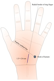
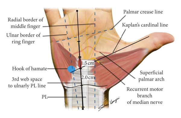
- The midpoint of the incision should be at the intersection of Kaplan cardinal line and radial border of 4th ring finger.
- The Kaplan cardinal line is a line drawn between the apex of the 1st web space to the pisiform.
- The incision should extend to the transverse palmar crease distally
- Don’t extend the proximal extent beyond the volar wrist crease.
- The midpoint of the incision should be at the intersection of Kaplan cardinal line and radial border of 4th ring finger.
- Dissection into the carpal tunnel
- Deepen the incision through the longitudinal fibers of the Palmar aponeurosis to expose the transverse fibers of the flexor retinaculum.
- Use a West retractor
- Incise the flexor retinaculum to expose the median nerve
- Pass a McDonald dissector below the flexor retinaculum while the remaining transverse fibers are divided.
- Ensure the proximal retinaculum has been released
- Flexor tendons
- If there is an associated synovitis affecting the flexor tendons - perform a flexor synovectomy.
- Close skin
Nerves at risk during a carpal tunnel release
- Palmar cutaneous branch of median nerve - enters palm superficial to retinaculum.
- Recurrent branch of median nerve - comes off median nerve distal to flexor retinaculum, curls proximally around distal border of retinaculum to supply the thenar eminence.