Section: Skin and soft tissue Sub-section: Peripheral nerve issues Curriculum: Curriculum, page 56
Ulnar Nerve entrapment
Elbow - Cubital tunnel syndrome
- Most common cause of ulnar nerve entrapment
- Compressive neuropathy of the ulnar nerve caused by anatomic compression in the medial elbow
- Positive Tinels sign over the cubital tunnel
Causes
- Entrapment
- Most common sites
- between the two heads of FCU/aponeurosis (most common site)
- within Arcade of Struthers (hiatus in medial intermuscular septum)
- between Osborne’s ligament and MCL
- Most common sites
- External compression
- Trauma
- Nerve compression, traction or friction - from leaning on the elbow or prolonged flexion.
- Joint pathology - osteophytes, arthritis, ganglia.
Clinical features
- Sensory symptoms in the ulnar nerve distribution including the dorsal and palmar cutaneous aspects
- Motor symptoms - weakness of the hand.
Wrist - Ulnar tunnel syndrome
- Less common
- Also referred to as Guyon’s canal syndrome
- Is ulnar neuropathy at the wrist where it passes through the Guyon’s canal.
Causes
- Trauma
- Lacerations.
- Repetitive use of tools in the workplace
- Use of a walker.
- Ganglia
Clinical features
-
Typically presents with hand weakness and sensory involvement.
-
The sensory involvement typically does not involve the distribution of the dorsal cutaneous nerve or palmer cutaneous branch as these are given off in the forearm.
-
The clawing of digits 4 and 5 is generally worst because of the unopposed action of flexor digitorum profundus (Ulnar paradox)
-
Guyons canal can be broken into 3 zones - depending on what zone is impinged, will depend on what symptoms the patients has
- Zone 1 - common branch of ulnar nerve - impingement results in sensory and motor symptoms.
- Zone 2 - impingement results in motor symptoms - deep motor branch supplies interossei, the 3rd and 4th lumbricals, and Opponens digiti minimi - paralysis causes mild hand clawing
- Zone 3 - impingement results in sensory symptom - superficial sensory terminal branch - ulnar border of the palm and then 1.5 digits on the palmar aspect an the tip
Examination of ulnar nerve
- Look:
- Evidence of claw hand
- Flexed 4th and 5th metacarpals with extended proximal and distal phalangeal joints.
- Hypothenar wasting
- Guttering between metacarpals
- Evidence of claw hand
- Sensation
- 3 territories
- dorsal cutaneous
- superficial terminal
- palmar cutaneous
- 3 territories
- Strength
- Test strength - flexion of 4th and 5th fingers (lumbricals)
- Test interossei - abduction of fingers
- Test adductor pollicis - patient holds paper between thumb and second finger (Froment’s test)
- Provocative tests
- Froments Test - hold paper between thumb and radial side of 2nd digit
- Sign presents after damage to the ulnar nerve, which innervates the adductor pollicis and interossei muscles, which provide adduction of the thumb and extension of the interphalangeal joint. The flexor pollicis longus (innervated by the median nerve), will substitute for the adductor pollicis (innervated by the ulnar nerve) and cause the thumb to go into hyperflexion.
- Elbow Flexion Test - flex elbow for 1 minute
- Numbness and tingling 4th and 5th digits
- Tinels Tap
- Can be performed at cubital tunnel and Guyon’s canal
- Causes numbness and tingling in ulnar nerve distribution
- Froments Test - hold paper between thumb and radial side of 2nd digit
- Ulnar paradox
- High lesion
- Proximal
- Prior to innervation of FDP to 4th and 5th digit
- Less clawing as action of FDP lost
- Low lesion
- Distal
- Distal to innervation of FDP to 4th and 5th digit
- More clawing as action of FDP remains - MCPJ hyperextension and flexion PIPJ and DIPJ for digits 4 and 5 possible
- Still get hypothenar wasting and loss of ADD and ABD
- High lesion
Management
- Ulnar tunnel syndrome
- Behavioural change:
- wrist brace
- Surgery
- Ulnar nerve decompression
- indicationsn
- severe symptoms that have failed nonoperative treatment
- indicationsn
- tendon transfers
- indications
- correction of clawed fingers
- loss of power pinch
- Wartenberg sign (abduction of small finger)
- indications
- Ulnar nerve decompression
- Behavioural change:
- Cubital tunnel syndrome
- Behavioral change:
- avoid leaning on elbow
- use of a soft foam elbow
- wraps of the elbow to limit flexion at nightime
- Surgery
- Indications
- When non-operative management fails
- Options
- in situ ulnar nerve decompression without transposition
- ulnar nerve decompression and anterior transposition
- medial epicondylectomy
- Indications
- Behavioral change:
Median Nerve injury - Pronator syndrome
-
Pronator Syndrome is a compressive neuropathy of the Median Nerve at the level of the elbow.
-
Diagnosis is made clinically with pain at the proximal volar forearm, sensory changes over the palmar cutaneous branch, and positive Tinel’s over the proximal volar forearm.
-
Treatment involves a prolonged nonoperative course, and rarely, surgical decompression.
-
Pathoanatomy
- 5 potential sites of entrapment include
- supracondylar process
- residual osseous structure on distal humerus present in 1% of population
- ligament of Struthers
- travels from tip of supracondylar process to medial epicondyle
- not to be confused with arcade of Struthers which is a site of ulnar compression neuropathy in cubital tunnel syndrome
- bicipital aponeurosis (a.k.a. lacertus fibrosus)
- between ulnar and humeral heads of pronator teres
- considered the most common site of entrapment
- FDS aponeurotic arch
- supracondylar process
- 5 potential sites of entrapment include
-
Associated conditions
- commonly associated with medial epicondylitis
-
The median nerve is a terminal branch of the brachial plexus, taking divisions from the lateral and medial cords to derive supply from C5-T1.
-
It supplies most of the flexor muscles of the forearm, but only the thenar muscles (FPL, OP, APB) and two lumbricals in the hand. 1/2LOAF
-
In the arm it is medial to the brachial artery but it crosses the ulnar artery just distal to the ACF to run down the midline of the forearm where it dives between the heads of pronator teres and deep to the arch of FDS.
-
Prior to this it supplies PL, PT, FCR, and FDS.
-
Its anterior interosseous branch is the nerve of the deep flexor compartment supplying the radial half of FDP, as well as PQ and FPL.
-
It enters the hand through the carpel tunnel where it may be compressed causing Carpal tunnel syndrome
-
Sensory supply of the median nerve is to the radial 3.5 digits on the entire palmar side and the tips of the dorsal digits.
-
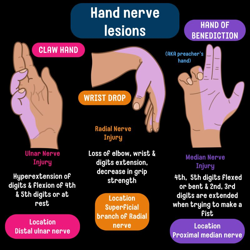
The Hand of Benediction occurs when a patient with a high median nerve injury (proximal to the elbow) attempts to make a fist, but the index and middle fingers remain extended, while the ring and little fingers flex.
-
Nonoperative
- rest, splinting, and NSAIDS for 3-6 months
- indications
- mild to moderate symptoms
- technique
- splint should avoid forearm rotation
- indications
- rest, splinting, and NSAIDS for 3-6 months
-
Operative - Decompressive surgery for pronator syndrome
- surgical decompression of median nerve
- indications
- only when nonoperative management fails for 3-6 months
- technique
- decompression of the median nerve at all 5 possible sites of compression
- outcomes
- of surgical decompression are variable
- 80% of patients having relief of symptoms
- of surgical decompression are variable
- indications
- surgical decompression of median nerve
Lateral cutaneous nerve entrapment
Intro
- The lateral femoral cutaneous nerve is a purely sensory nerve that is susceptible to compression.
- Meralgia Paresthetica
Risk factors
- Obesity
- Diabetes mellitus
- Older age
Causes
- Body habitus - large abdomen
- Pregnancy
- Increased intra-abdominal pressure due to ascites
- Tight belts
- Compression from a wallet.
- Prolonged leaning of a thigh against a bench or table.
- Carrying heavy objects.
- Long distance walking
- Groin trauma
- Surgery - inguinal hernia repair, iliac bone harvesting, aorto-bifemoral bypass, spine surgery
Anatomy
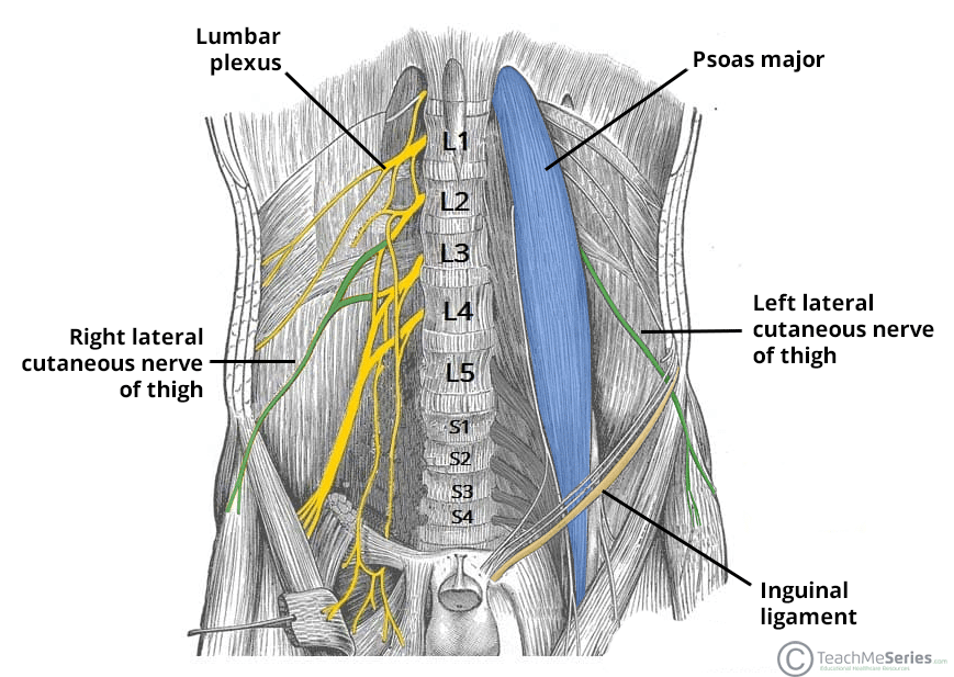
- The lateral femoral cutaneous branches off the lumbar plexus and conveys fibers from L2 and L3 nerve roots.
- The nerve courses through the pelvis running adjacent to the lateral edge of psoas
- It enters the leg underneath or through the inguinal ligament, medial to the ASIS - in this location, entrapment can occur.
Clinical features
Symptoms
- Pain, paresthesia, and numbness over the upper outer thigh
Signs
- Loss of light touch or pinprick sensation of the upper outer thigh.
DDx
- L3/L4 radiculopathy
- Femoral neuropathy - passes under the inguinal ligament medial to the lateral femoral cutaneous and innervates the upper medial aspect of the thigh. You will also get weakness of the quadriceps (knee extension)
Treatment
- Is self-limiting in 90% of cases.
Conservative treatment
- Weight loss
- Avoid pressure to groin.
- Patient reassurance that it does not represent a back problem
Persistent symptoms
- Gabapentin.
- Local nerve block.
Surgical
- Decompression
- Nerve transection - last resort.
Radial Nerve neuropathy
Anatomy
- C5-T1 brachial plexus posterior cord.
- Supplies extensor arm and forearm compartments
- Supplies sensation to dorsal hand, forearm and upper arm.
Injury
- Saturday night palsy - arm leaning over side of chair, compresses it in axilla.
- Humeral fracture
Symptoms and signs
- Wrist drop
- Loss of elbow extension
- Loss of wrist extension
- Loss of MCP extension
- Preservation of extension at DIPJ and PIPJ due to action of DAB and PAD, lumbricals.
Other injuries
Peripheral nerve injuries associated with GA
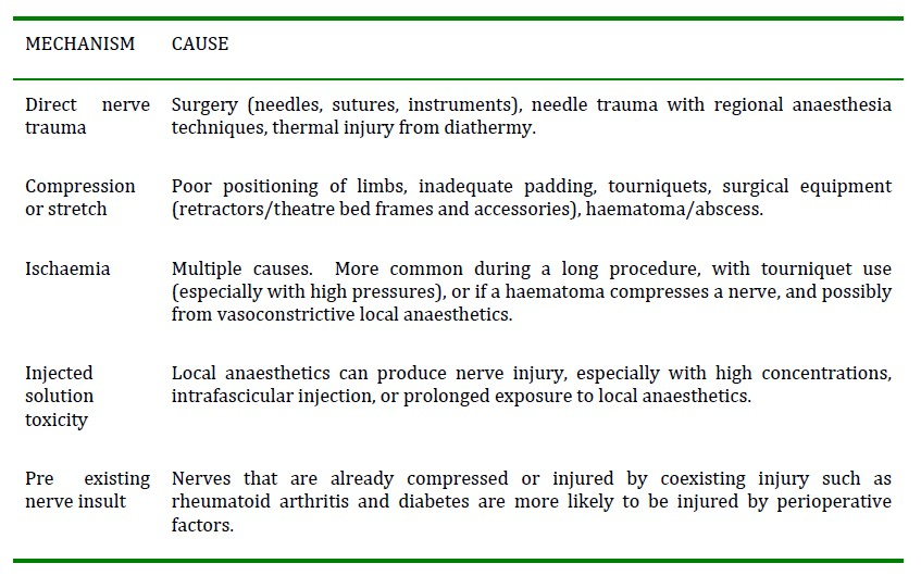
Mechanism
Common peripheral nerve injuries
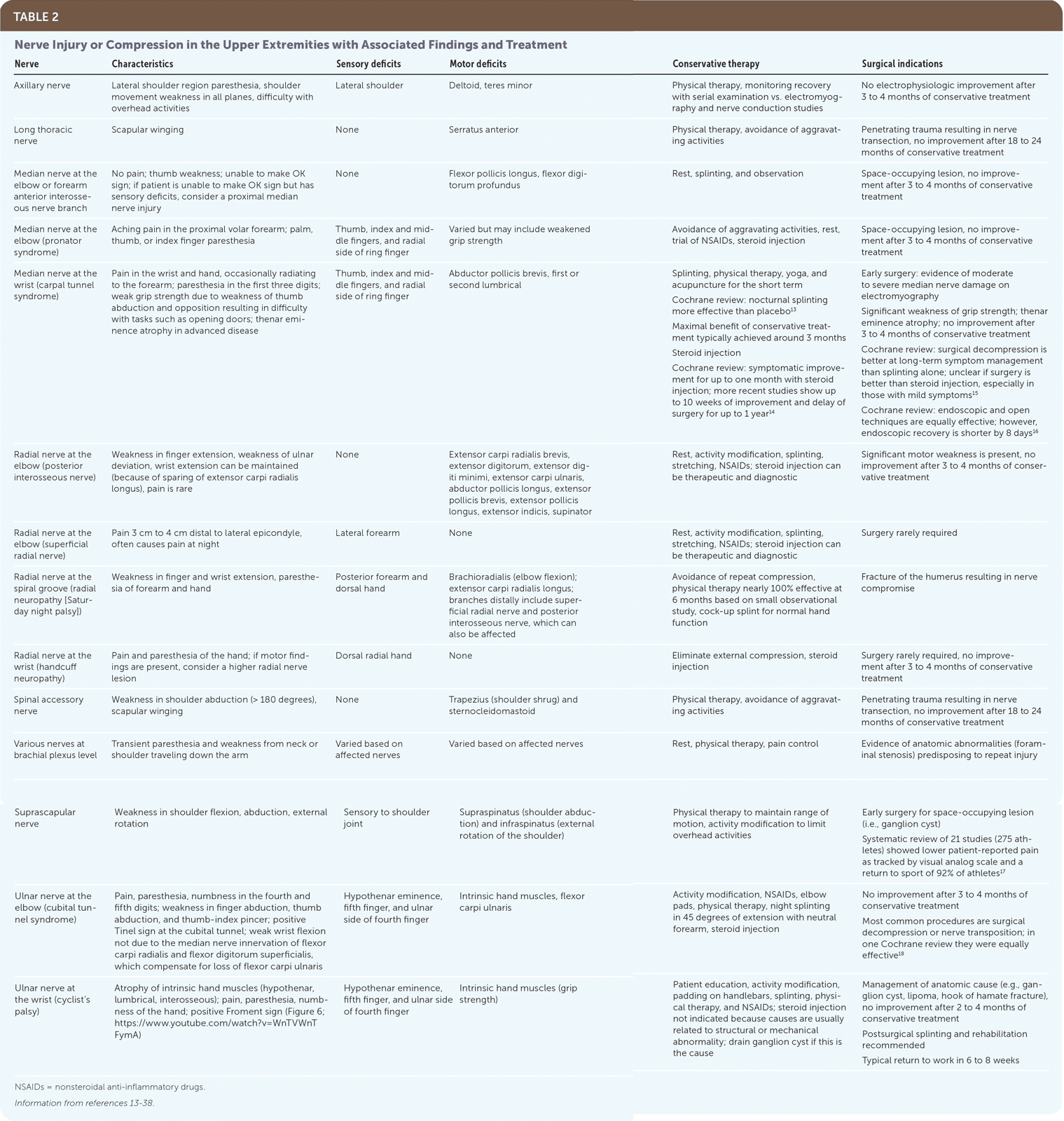
Ulnar neuropathy
- Usually occurs at the superficial condylar groove of the elbow
- Most common peripheral nerve injury.
- Vulnerable to compression against the operating table, especially with forearm extension and pronation.
- Extreme elbow flexion with the arm across the chest can lead to stretch injury of the ulnar nerve
Brachial plexus neuropathy
- The most common causes of brachial plexus neuropathy are stretch, compression or direct injury (via surgical or regional techniques).
- Brachial plexus stretch can be caused by arm abduction, external rotation and posterior shoulder displacement.
- This can occur with a supine patient when the neck is flexed laterally, along with an abducted externally rotated arm position.
- Extreme abduction of the arm, (with hands above the head) also stretches the brachial plexus.
- Compression of the brachial plexus has been associated with sternotomy and sternal retraction.
- Compression can also occur in the lateral position when the plexus is compressed between the thorax and the humeral head.
- The long thoracic nerve can be damaged in brachial plexus PPNI, causing winging of the scapula.
Lumbosacral nerve root neuropathy
- Association with a regional technique
- Majority of these are associated with central neuraxial approaches
Common peroneal nerve
- This is the most frequent of the lower limb nerve injuries.
- The common peroneal nerve is at risk of injury in the lithotomy position, where it can be compressed by the head of the fibula, or in the lateral position where it can be compressed between the fibula and the operating table.
Radial Nerve
- Radial nerve injury occurs most commonly due to compression of the radial nerve between the operating table and the humerus.
- Radial nerve injury can also occur when a patient is in the lateral position with their upper arm abducted beyond ninety degrees and suspended from a support.
Sciatic nerve
- The sciatic nerve is more at risk with very thin patients, long procedures and hard operating surfaces.
- Elevation of the opposite buttock (such as for some hip surgery) increases the risk further.
- Excessive stretch of the sciatic nerve can occur when the lithotomy position is used and extreme external rotation of the thigh is applied.
- Sciatic nerve injury has been reported post coronary artery bypass graft, possibly from prolonged nerve pressure along with low perfusion pressure.