- Roots:
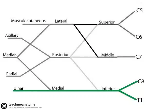
- C8 and T1 from Brachial Plexus
- Motor Functions
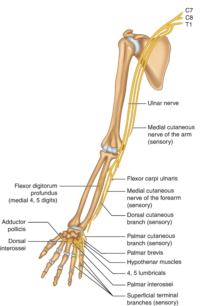
- Muscles of the hand (apart from the thenar muscles and two lateral lumbricals)
- Flexor carpi ulnaris
- Medial half of flexor digitorum profundus.
- Sensory Functions
- Innervates the anterior and posterior surfaces of the medial one and half fingers, and associated palm area.
- Innervates the anterior and posterior surfaces of the medial one and half fingers, and associated palm area.
Ulnar Nerve in the Axilla
- Fibers from C7, C8, T1
- Largest branch of Medial Cord
- Runs down between Axillary Artery and Axillary Vein
- The most posterior of the structures which run down the medial side of the flexor compartment of the arm
Ulnar Nerve in the Arm
- Upper arm
- In the upper arm the ulnar nerve lies in close proximity to the brachial artery and median nerve
- This is located in the medial aspect of the arm between the anterior and posterior compartments
- It has no motor or sensory branches above the elbow.
- In the upper arm the ulnar nerve lies in close proximity to the brachial artery and median nerve
- Pierces
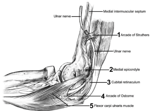
- The Medial Intermuscular Septum in lower third of arm
- Accompanied by Superior Ulnar Collateral Artery and a branch of Radial Nerve to Medial Head of Triceps
- The Medial Intermuscular Septum in lower third of arm
- Elbow
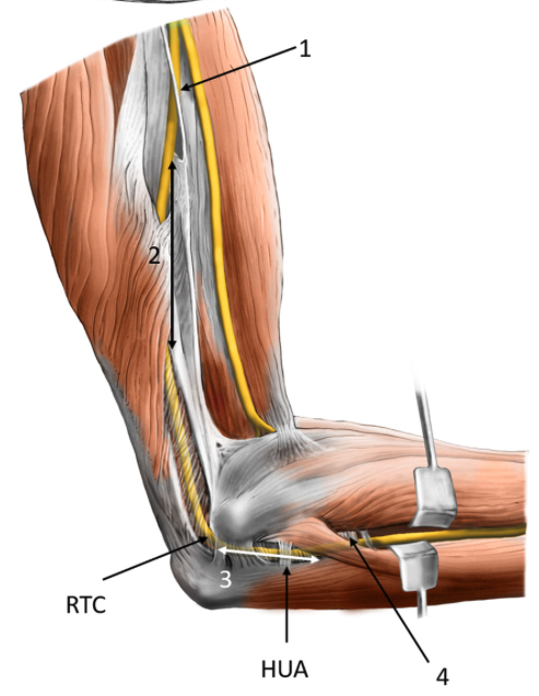
- Lies in the groove behind the retrocondylar groove behind the Medial Epicondyle of Humerus
- Palpable in this location
- Courses through the lower part of extensor compartment and disappears into forearm by passing between Humeral and Ulnar heads of origin of Flexor Carpi Ulnaris
- It passes under the aponeurotic arch of the flexor carpi ulnaris (called the humero-ulnar arcade) - this is an important landmark because entrapment can occur here (see the second picture)
- Lies in contact with the bone in the groove behind base of Medial Epicondyle
- Lies against the Medial Ligament of the Elbow Joint – which it supplies
- Lies in the groove behind the retrocondylar groove behind the Medial Epicondyle of Humerus
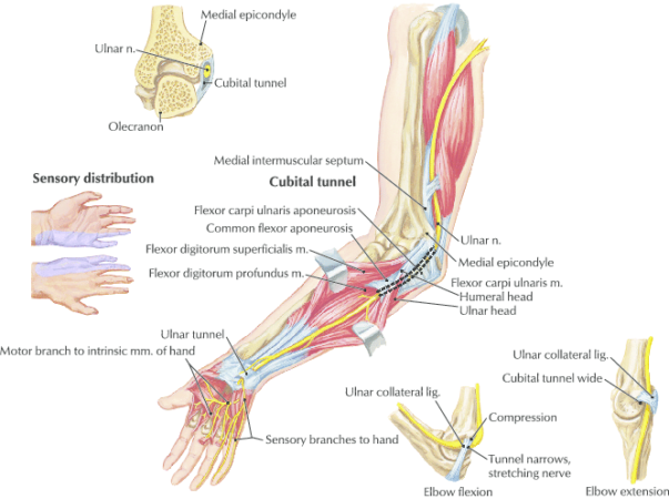
Ulnar Nerve in the Forearm
- Lies under cover of flattened aponeurosis of Flexor Carpi Ulnaris
- Ulnar Artery on its radial side
- Neurovascular bundle lies on Flexor Digitorum Profundus
- Supplies:
- Flexor Carpi Ulnaris
- Ulnar Half of Flexor Digitorum Profundus
- Emerges from behind tendon of Flexor Carpi Ulnaris proximal to the wrist
- Passes across the front of the Flexor Retinaculum lateral to the Pisiform bone in the hand
- Branches
- Gives off Dorsal branch before emerging
- Dorsal branch passes medially between tendon of Flexor Carpi Ulnaris and lower end of Ulna
- Supplies: Dorsum of the Hand and Ulnar one and a half fingers proximal to nail beds
- Dorsal branch passes medially between tendon of Flexor Carpi Ulnaris and lower end of Ulna
- Gives off Palmar Cutaneous Branch which pierces deep fascia proximal to Flexor Retinaculum
- Supplies: Skin of Hypothenar Eminence
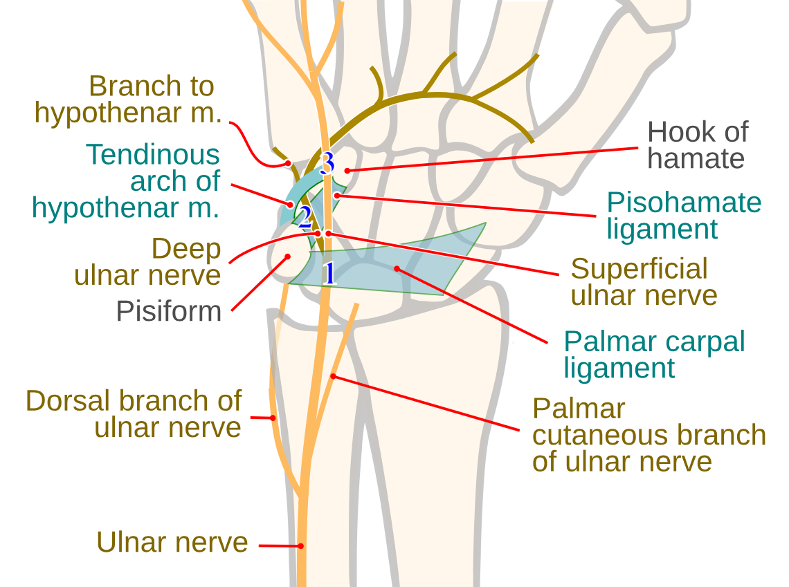
- Supplies: Skin of Hypothenar Eminence
- Gives off Dorsal branch before emerging
Ulnar Nerve in the Hand
- Wrist
- At the wrist, the ulnar nerve passes through Guyon’s canal (along with the ulnar artery)
- The floor of the canal is formed by the transverse carpal ligament and pisohamate ligament.
- The roof consists of the palmar fascia and palmar brevis.
- Guyons canal can be broken into 3 zones - depending on what zone is impinged, will depend on what symptoms the patients has .
- Zone 1 - common branch of ulnar nerve - impingement results in sensory and motor symptoms.
- Zone 2 - impingement results in motor symptoms - deep motor branch supplies interossei, the 3rd and 4th lumbricals, and Opponens digiti minimi - paralysis causes mild hand clawing
- Zone 3 - impingement results in sensory symptom - superficial sensory terminal branch - ulnar border of the palm and then 1.5 digits on the palmar aspect an the tip.
- At the wrist, the ulnar nerve passes through Guyon’s canal (along with the ulnar artery)
- Branches
- Divides on the Flexor Retinaculum alongside Pisiform bone
- Superficial Branch and Deep Branch
- Superficial Branch:
- Runs beneath Palmaris Brevis
- Distributed by 2 digital branches to the ulnar one and a half fingers, including nail beds and skin on dorsum not supplied by dorsal branch
- Supplies: Palmaris Brevis
- Deep Branch:
- Runs deeply between Abductor Digiti Minimi and Flexor Digiti Minimi and then through Opponens Digiti Minimi
- Supplies: All 3 Hypothenar Muscles (Abductor Digiti Minimi, Flexor Digiti Minimi, Opponens Digiti Minimi)
- Grooves the distal border of the Hook of Hamate
- Crosses the palm in the concavity of the Deep Palmar Arch
- Supplies: Two Ulnar Lumbricals, all Palmar and Dorsal Interossei, and Adductor Pollicis
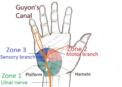
- Divides on the Flexor Retinaculum alongside Pisiform bone
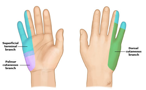
Branches
- Forearm
- Articular branches
- Elbow joint
- Muscular branches
- Flexor carpi ulnaris
- Medial half of the flexor digitorum profundus
- Palmar cutaneous branch
- Arises from the ulnar nerve around the mid-portion of the forearm.
- Runs along the ulnar artery to the distal forearm, where it perforates the deep fascia.
- Sensory innervation to the skin at base of the medial palm
- Dorsal cutaneous branch
- Emerges in the distal forearm, proximal to the wrist.
- Courses posteriorly, deep to the flexor carpi ulnaris and perforates the deep fascia to enter the medial aspect of the dorsum of hand.
- It divides into two or three dorsal digital nerves to supply sensation to the skin on the medial side of the dorsum of hand as well as the proximal regions of the medial 1½ digit (not the tips)
- Articular branches
- Hand
- Superficial branch of ulnar nerve
- Sensory innervation to the anterior aspect of the ulnar 1½ digits (little finger and half of the ring finger) and medial palmar skin.
- Tips of both fingers palmer and volar
- Motor innervation to the palmaris brevis muscle in the hypothenar region of the hand.
- Arises from the ulnar nerve just distal to the pisiform bone and often divides into two palmar digital nerves.
- Sensory innervation to the anterior aspect of the ulnar 1½ digits (little finger and half of the ring finger) and medial palmar skin.
- Deep branch of the ulnar nerve
- Emerges from the same region of the nerve as the superficial branch
- Courses between the muscles of the hypothenar region into the deep aspect of the plam, alongside the deep palmar branch of the ulnar artery.
- Provides motor innervation to the vast majority of hand muscles.
- Superficial branch of ulnar nerve
Brief summary
- Origin:
- C8 and T1 from Brachial Plexus
- Continuation of the medial cord
- Motor Functions
- Muscles of the hand (apart from the thenar muscles and two lateral lumbricals)
- Flexor carpi ulnaris
- Medial half of flexor digitorum profundus.
- Sensory Functions
- Innervates the anterior and posterior surfaces of the medial one and half fingers, and associated palm area. Course
- Axilla
- Runs down between Axillary Artery and Axillary Vein
- Arm
- Run with brachial artery and median nerve on the medial aspect of the arm between the anterior and posterior compartments
- Pierces the Medial Intermuscular Septum in lower third of arm to enter the posterior compartment
- Elbow
- Lies in the retrocondylar groove behind the Medial Epicondyle of Humerus
- Forearm
- Passing between the two heads of Flexor Carpi Ulnaris
- Lies under the Flexor Carpi Ulnaris and on the Flexor Digitorum Profundus - supplies both
- Ulnar Artery on its radial side
- Supplies:
- Flexor Carpi Ulnaris
- Ulnar Half of Flexor Digitorum Profundus
- Emerges from behind tendon of Flexor Carpi Ulnaris proximal to the wrist
- Branches
- Dorsal cutaneous
- Palmer cutaneous
- Wrist
- Passes across the front of the Flexor Retinaculum lateral to the Pisiform bone in the hand
- Divides on the Flexor Retinaculum alongside Pisiform bone
- Superficial Branch - sensory
- Deep Branch - muscular