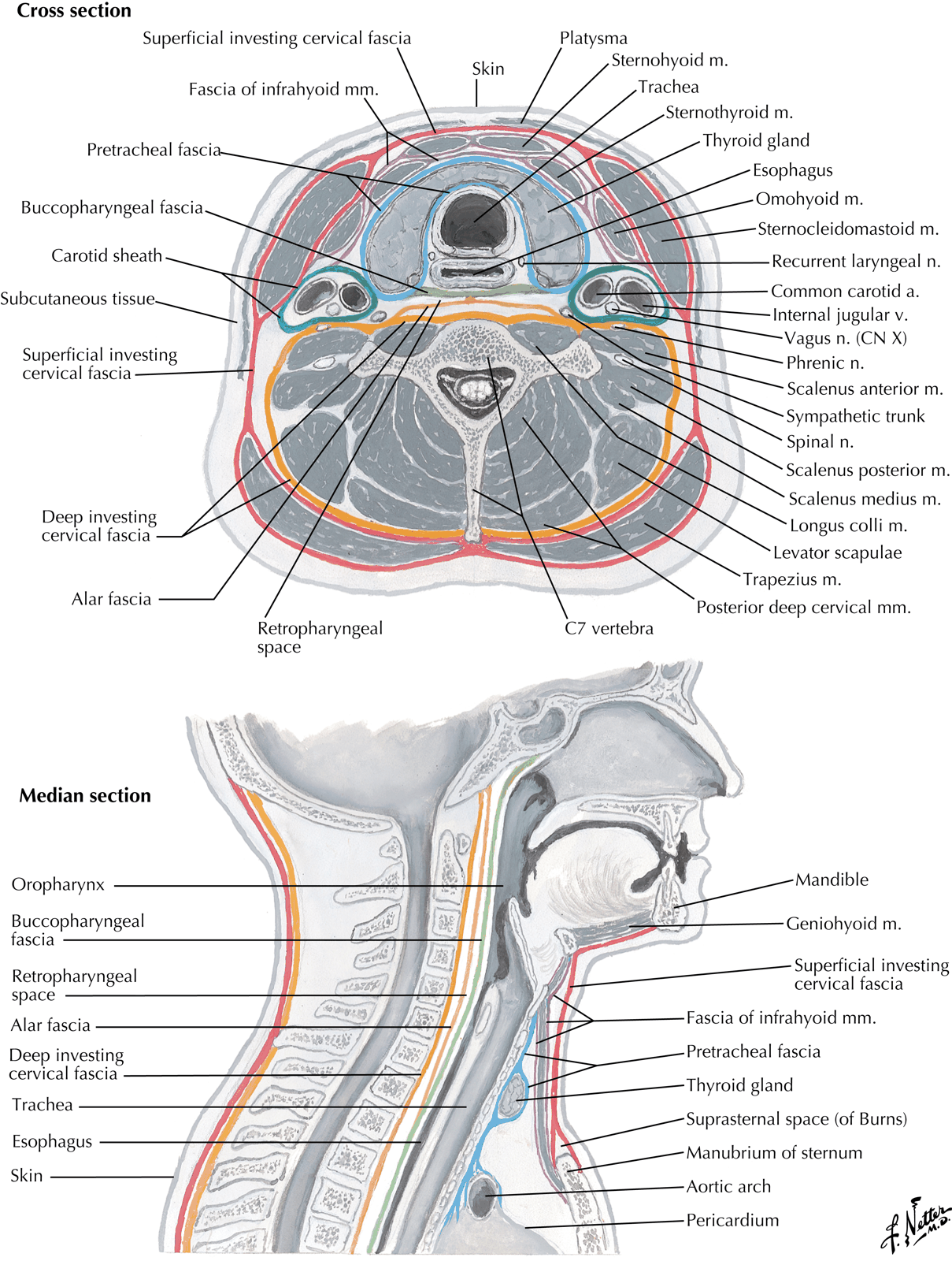
Superficial cervical fascia
- The contents of the superficial cervical fascia will vary depending upon location but include:
- Platysma (and cervical branch of facial nerve that innervate the muscle)
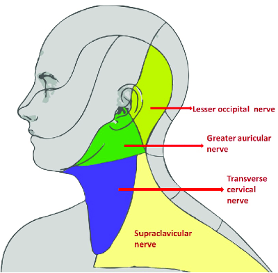
- Cutaneous nerves of the cervical plexus:
- Lesser occipital
- Great auricular
- Transverse cervical (largest sensory contribution)
- Supraclavicular
- Deep to the superficial cervical fascia is the investing (or superficial) layer of deep cervical fascia
Superficial Structures
Platysma
- Lies superficial to Investing Layer of Deep Cervical Fascia
- Extends from fascia over upper parts of Pectoralis Major and Deltoid to lower border of Mandible
- Some fibres extend into face blending with fascial expression muscles
- Covers External and Anterior Jugular Veins
- Separated below but converge above towards midline just beneath chin
- Nerve Supply:
- Cervical Branch of Facial Nerve
- Afferent (proprioceptive) fibres run with Transverse Cervical Nerve
- Action:
- Involved in facial expression
- May assist in mouth opening
Anterior Jugular Veins
- Commence beneath chin and pass downwards
- Side by side beneath Platysma
- Reaches Suprasternal region
- Pierce Deep Fascia and come to lie in Suprasternal Space (often connected by a short anastomotic vein)
- Angles laterally to pass deep to Sternocleidomastoid but superficial to Strap muscles
- Opens into the External Jugular Vein near its termination
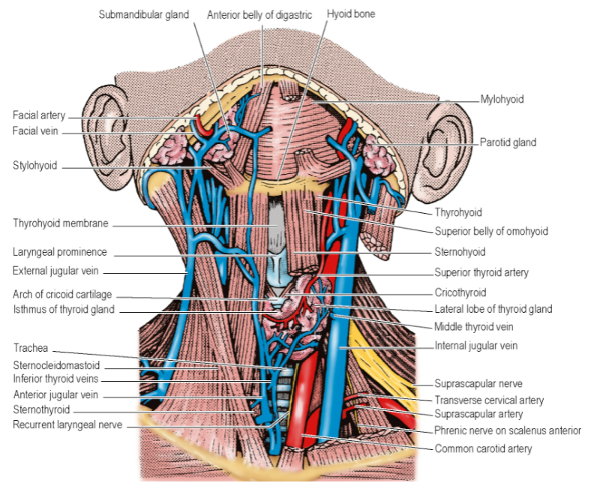
Deep Cervical Fascia
- Four parts:
- Superficial (Investing) Layer
- Pretracheal Fascia
- Prevertebral Fascia
- Carotid Sheath
- Investing Layer
- Surrounds neck like a collar
- Splits around Sternocleidomastoid and Trapezius
- NB: Investing layer does NOT enclose the infra-hyoid muscles
- Posteriorly, blends with Ligamentum Nuchae (attached to spines of Cervical Vertebrae)
- Anteriorly – attached to Hyoid Bone
- Above – attached to lower border of the Mandible
- Also attached to:
- Superior Nuchal Line
- External Occipital Protuberance at Base of Skull
- Encloses Parotid Gland between the Angle of the Mandible and the Tip of the Mastoid Process
-
Superficial part:
- Extends superiorly as the Parotidomasseteric Fascia
- Reaches Zygomatic Arch
-
Deep part:
- Extends to Base of Skull
- Forms the Stylomandibular Ligament between the Styloid Process and Angle of Mandible
-
Below:
- Investing Layer attached to Spine and Acromion of Scapula and Clavicle with Trapezius
- Attached to Clavicle and Manubrium with Sternocleidomastoid
- In between these muscles, attached to both Clavicles and Jugular (Suprasternal) Notch by 2 layers into which it splits a short distance above them
- Suprasternal Space:
- Formed between layers of Investing Layer attached to anterior and posterior borders of Jugular Notch
- Contents include:
- Lower parts of the Anterior Jugular Veins
- An anastomotic arch between them
- Sternal Heads of Sternocleidomastoids
- Sometimes a Lymph Node
- In the middle third of the Clavicle – the deeper of the two layers splits around Inferior Belly of Omohyoid – forms a fascial sling keeping the muscle belly low down in the neck
- Two layers pierced by External Jugular Vein
- Prevertebral Fascia
- Lies in front of the Prevertebral muscles
- Extends from Base of Skull downwards to blend with Anterior Longitudinal Ligament of T4 Vertebral Body in the Superior Mediastinum
- Thus, infections behind the Prevertebral Fascia imprisoned in the Superior Mediastinum in front of the vertebral bodies
- Anterior to Longus Capitis, Rectus Capitis Lateralis, Longus Colli
- Extends sideways across Scalenus Anterior, Scalenus Medius, Levator Scapulae Muscles
- Gets thinner further out laterally
- Fades under cover of anterior border of Trapezius
- Covers muscles that form the floor of the Posterior Triangle of the Neck and all Cervical Nerve Roots
- Thus, Cervical Plexus and Trunks of Brachial Plexus lie deep to it
- Lymph Nodes of Posterior Triangle and Accessory Nerve lie superficial to it
- Cervical Sympathetic Trunk does NOT lie deep to the Prevertebral Fascia
- 3rd part of Subclavian Artery – deep to Prevertebral Fascia
- Prevertebral Fascia becomes prolonged over the Subclavian Artery and the Brachial Plexus below the Clavicle as the Axillary Sheath
- Does not invest the Subclavian or Axillary Vein
- These lie in loose areolar tissue anterior to it
- Free to dilate during times of increased venous return from the upper limb
- Does not invest the Subclavian or Axillary Vein
- Prevertebral Fascia pierced by 4 cutaneous branches of the Cervical Plexus
- Great Auricular Nerve
- Lesser Occipital Nerve
- Transverse Cervical Nerve
- Supraclavicular Nerve
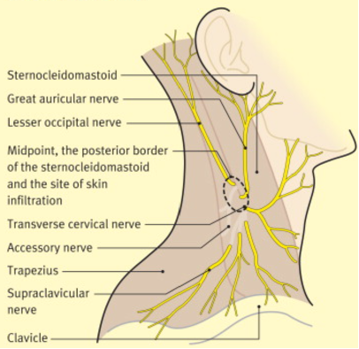
- Pretracheal Fascia
- Deep to Infrahyoid Strap muscles (Sternothyroid, Sternohyoid, Omohyoid)
- Upward attachment limited by respective attachments of those muscles
- I.e. Body of Hyoid Bone and Oblique Line of Thyroid Cartilage
- Splits to enclose Thyroid Gland
- Not adherent to Thyroid except:
- At back of Isthmus – attached to 2nd, 3rd and 4th Tracheal Rings
- Laterally – fuses with front of Carotid Sheath on deep surface of Sternocleidomastoid
- Inferiorly – passes behind Brachiocephalic Veins to blend with Adventitia of Aortic Arch and Fibrous Pericardium
- Thus, neck infections in front of the Pretracheal Fascia is directed into the anterior mediastinum
- Part of Cervical Visceral Fascia surrounding Pharynx, Oesophagus, Larynx and Trachea
- Carotid Sheath
- Feltwork of areolar tissue
- Surrounds Common and Internal Carotid Arteries, Internal Jugular Vein, Vagus Nerve, and Deep Cervical Lymph Nodes
- Common Carotid Artery is medial to the Internal Jugular Vein
- Vagus Nerve deeply place between the two vessels
- Common Carotid Artery is medial to the Internal Jugular Vein
- Thin where it overlies Internal Jugular Vein – allows venous dilatation
- Attached to Base of Skull at margins of Carotid Canal and Jugular Fossa
- Continued downwards to blend with Adventitia of Aortic Arch
- Anteriorly – lower part of Sheath fuses with fascia on deep surface of Sternocleidomastoid
- Blends with Pretracheal Fascia where they lie alongside
- Posteriorly – minimum loose areolar tissue between it and Prevertebral Fascia
- Cervical Sympathetic Trunk lies in this space anterior to Prevertebral Fascia